Shoulder pain causes often originate within the soft tissues of the rotator cuff or subacromial space, structures that standard X-rays cannot visualize due to their reliance on ionizing radiation which only registers dense, calcium-rich bone structures. Consequently, identifying the root driver of chronic shoulder pain requires high-resolution soft tissue imaging to detect microscopic tendon fraying, bursal hypertrophy, or mechanical impingement. A thorough clinical assessment combined with advanced imaging allows us to map the precise pathology and design a safe, functional rehabilitation program.
In my clinical practice in Vigan, Ilocos Sur, I regularly consult with patients—ranging from local farmers carrying heavy sacks of rice (paggamas) to tricycle drivers navigating rough provincial roads—who complain of chronic, nagging shoulder pain. They often describe this as a deep ngawit (heaviness) or a sharp kirot (pain) that occurs during overhead activities. A common source of frustration for these patients is having a "normal" shoulder X-ray despite suffering from severe, disabling pain. It is vital to explain to patients that a standard X-ray is designed to rule out bone-related pathologies like fractures, severe glenohumeral osteoarthritis, or subacromial bone spurs. It cannot, however, register the structural integrity of the tendons, ligaments, or the subacromial bursa, where the vast majority of shoulder pain generators actually reside.
Contrarian Insight 1: A "normal" shoulder X-ray is frequently misinterpreted by patients and some primary care providers as a sign that no structural damage exists, leading to a false sense of security. This misunderstanding often delays critical diagnostic imaging, prompting patients to rely on chronic oral non-steroidal anti-inflammatory drugs (NSAIDs) that mask progressive soft-tissue degeneration and potentially accelerate tendon wear by suppressing necessary physiological remodeling.
To understand why this soft tissue pain is so disabling, we must look at the biomechanics of the shoulder. The glenohumeral joint is a ball-and-socket joint characterized by a large humeral head and a shallow glenoid cavity—much like a golf ball sitting on a tee. This anatomical design allows for an extraordinary range of motion, but it sacrifices inherent bony stability. To compensate, the joint relies on a dynamic stabilizer network known as the rotator cuff, alongside the stabilizing negative pressure of the glenoid labrum. When any of these soft tissue structures are injured, inflamed, or frayed, the dynamic stability of the shoulder is compromised. This results in micro-instability and mechanical friction that manifests as sharp pain when you attempt to lift your arm sideways, reach behind your back, or perform manual work.
Rotator cuff tears represent structural compromises in one or more of the four stabilizing shoulder tendons—primarily the supraspinatus—resulting from acute traction forces or progressive, age-related hypovascular degeneration. These structural defects present clinically as localized shoulder pain, nocturnal sleep disruption when lying on the affected joint, and mechanical catching during arm elevation. Identifying the exact tendon involved and the depth of the tear is critical, as these details directly dictate our rehabilitation approach and define the boundaries of conservative management.
The rotator cuff is composed of four distinct muscle-tendon units that work in structural harmony:
We classify rotator cuff tears by their structural depth and size. Partial-Thickness Rotator Cuff Tears (PTRCT) involve disruption of some tendon fibers while the remainder remain anchored to the bone. These are further sub-classified based on their location: articular-sided (facing the joint cavity), bursal-sided (facing the subacromial bursa), or intrasubstance (within the tendon belly). In contrast, Full-Thickness Rotator Cuff Tears represent a complete discontinuity of the tendon fibers from the bone footprint, which can range from small pinholes to massive retracted tears where the tendon has pulled back several centimeters from its insertion site.
Contrarian Insight 2: There is a significant mismatch between the severity of shoulder pain and the actual size of a rotator cuff tear. In clinical practice, a small, highly inflamed bursal-sided partial-thickness tear can generate intense, agonizing kirot and severe functional limitation due to chemical nociceptor activation in the highly vascularized bursa. Conversely, an elderly patient may present with a chronic, massive full-thickness supraspinatus tear and experience only mild ngawit or compensation-induced fatigue because their surrounding muscles have slowly adapted to stabilize the joint biomechanically over several years.
In the clinic, patients experiencing a rotator cuff tear typically present with a distinct cluster of symptoms. The hallmark symptom is intense nocturnal pain, particularly when rolling onto the affected shoulder. This pain is driven by increased intraosseous pressure and inflammatory fluid accumulation in the subacromial space when the joint is compressed. Patients also report a "painful arc"—a sharp catching or sumasabit sensation felt specifically when actively abducting the arm between 60 degrees and 120 degrees. When the arm is held below or lifted above this range, the pain often decreases. In chronic cases that have been neglected or treated only with traditional hilot (massage), we often observe pagliit ng balikat (atrophy of the supraspinatus and infraspinatus muscles), which presents as visible hollowing of the shoulder blade and profound weakness during arm elevation.
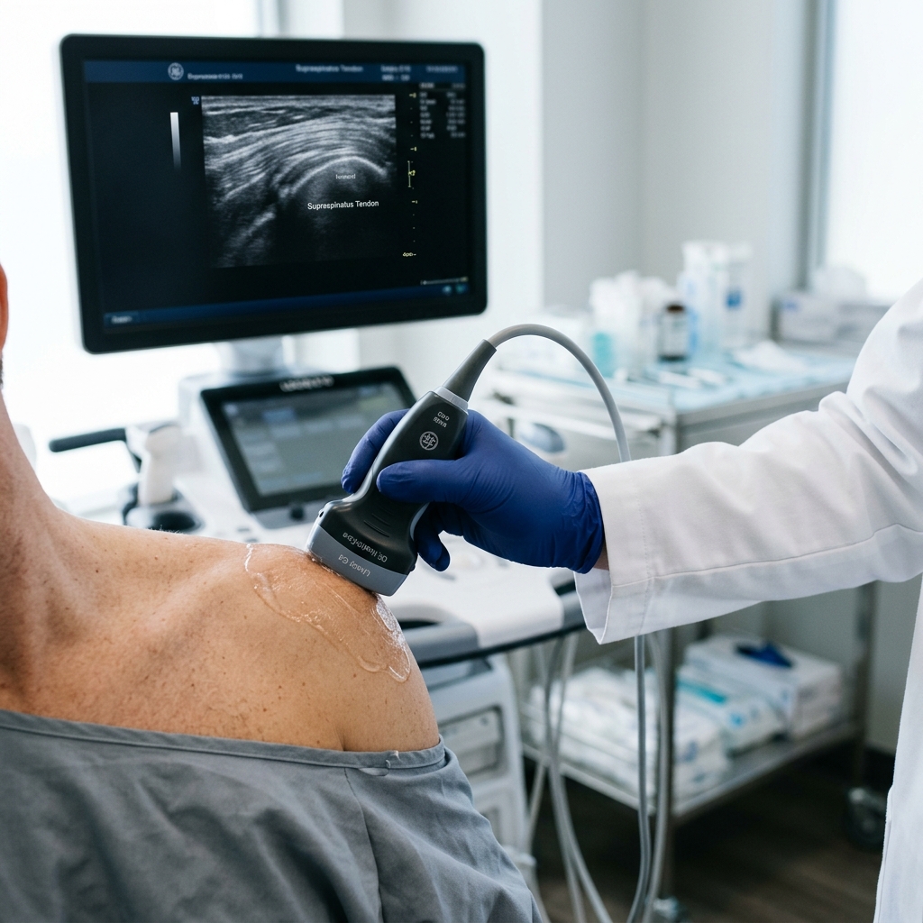
Musculoskeletal (MSK) ultrasound and magnetic resonance imaging (MRI) serve as the primary diagnostic modalities for rotator cuff pathology, with ultrasound offering real-time dynamic testing during joint movement and MRI providing high-resolution static evaluation of deep intra-articular structures. For superficial tendon and bursa assessments, clinical studies demonstrate that MSK ultrasound matches the diagnostic accuracy of MRI. Choosing between these modalities depends on cost, local availability, operator skill, and the specific clinical questions that need answering.
To appreciate the value of MSK ultrasound, we must contrast it directly with magnetic resonance imaging (MRI). An MRI requires the patient to lie completely still inside a narrow, noisy tube for 30 to 45 minutes. It captures static, high-resolution cross-sectional slices of the shoulder joint. MRI remains the gold standard for evaluating deep intra-articular structures, such as labral tears (SLAP lesions), bone marrow edema, occult fractures, and deep joint pathology. However, its static nature means it cannot show how the tendons and bones interact during movement. Musculoskeletal ultrasound, on the other hand, is a dynamic exam. Using a high-frequency linear probe (typically 10 to 18 MHz), the physician can directly visualize the rotator cuff tendons while actively moving the patient's arm. This allows us to observe subacromial impingement, dynamic snapping of the biceps tendon, or temporary subluxation in real-time as the patient reproduces the specific movements that trigger their pain.
A landmark systematic review and meta-analysis comparing the two modalities demonstrated that for experienced operators, MSK ultrasound has a diagnostic accuracy equivalent to MRI, with a median diagnostic accuracy of 0.93 (93%) for full-thickness supraspinatus tears and 0.81 (81%) for partial-thickness tears (Farooqi et al., 2021; PMID: 34660823). More recently, a clinical study evaluating diagnostic performance confirmed that ultrasound exhibits high sensitivity (91.2%) and specificity (81.8%) for identifying rotator cuff tears, validating its role as a primary diagnostic tool (Ganesh et al., 2024; PMID: 39350873). The major limitation of ultrasound is that it is highly operator-dependent; it requires a specialist with extensive training in musculoskeletal anatomy and dynamic scanning techniques to ensure accurate interpretation and avoid false-negative results.
Contrarian Insight 3: While MRI is widely regarded as the definitive diagnostic standard, static scans frequently identify age-related asymptomatic structural anomalies—such as mild labral fraying, minimal cartilage thinning, or asymptomatic partial tendon tears—in up to 50% of pain-free individuals over the age of forty. Relying solely on static MRI findings without dynamic clinical correlation can lead to diagnostic errors, attributing the patient's pain to a benign structural variant and potentially steering them toward unnecessary, invasive surgeries that fail to address the actual dynamic pain driver.
In the context of the Philippine healthcare system, particularly for patients in Northern Luzon, practical factors like travel and cost are critical components of the diagnostic plan. An MRI is a scarce resource in provincial areas, often requiring patients to travel 2 to 4 hours to larger cities like San Fernando or Metro Manila, with out-of-pocket costs ranging from ₱10,000 to ₱15,000. In contrast, MSK ultrasound can be performed directly in the Vigan clinic during the initial consultation. It is significantly more affordable, carries zero radiation risk, and permits immediate, hands-on discussion of the scan results with the patient, bridging the gap between imaging findings and the clinical rehabilitation plan.
Non-surgical treatment for rotator cuff disorders requires a structured sequencing of interventions, beginning with joint preservation and anti-inflammatory control, followed by guided mechanical loading to stimulate tendon repair. For partial-thickness tears, regenerative procedures like ultrasound-guided atelocollagen injections or platelet-rich plasma (PRP) therapy provide a structural matrix to accelerate tissue recovery. This staged approach ensures that we do not merely suppress pain symptoms temporarily, but actively promote tissue healing and restore long-term joint function.
When sequencing treatments, we must avoid the temptation to jump immediately to the strongest intervention or rely on repeated corticosteroid injections. While steroid shots are highly effective at extinguishing acute, chemical inflammation, they do not repair structural tears. In fact, repetitive steroid use can inhibit collagen synthesis, thins the tendon matrix, and increases the risk of tear progression. For patients with partial-thickness rotator cuff tears, our clinical focus has shifted toward regenerative medicine using ultrasound-guided atelocollagen or PRP injections to support the body's natural healing cascade. Atelocollagen, a highly purified type I collagen, acts as a sterile scaffold that recruits local cells to the injury site, promoting tissue regeneration while preserving the mechanical properties of the tendon during the recovery phase.
To implement this effectively, we apply strict clinical reasoning to define the targeted patient profile and monitor their progress:
Contrarian Insight 4: The common assumption that complete pain relief is the primary metric of successful treatment is clinically incomplete. Pain is an unreliable indicator of tissue healing; a tendon can become completely pain-free following rest or anti-inflammatory therapy while remaining structurally weak and prone to re-injury. True recovery is defined by the restoration of tissue load tolerance, muscle balance, and movement confidence, which can only be achieved through progressive, active loading rather than passive treatments alone.
Phased shoulder rehabilitation focuses on rebuilding rotator cuff load tolerance, restoring scapular stabilization, and re-establishing normal glenohumeral biomechanics through progressive mechanical loading. The rehabilitation program transitions from isometric muscle activation to dynamic resistance exercises, utilizing symptom-guided thresholds to prevent re-injury or persistent tendon inflammation. Rather than forcing the shoulder through painful movements, we dose the exercises based on the joint's current tissue irritability level.
The rehabilitation pathway is divided into three distinct clinical phases, mapped in the table below:
| Rehab Phase | Tissue Irritability & Symptoms | Clinical Targets & Exercise Dose |
|---|---|---|
| Phase 1: Protection & Activation | High irritability. Pain >7/10 at rest, severe night pain, unable to sleep, active elevation highly limited by sharp kirot. | Pain modulation, gentle passive range of motion (PROM) within pain-free limits, and sub-maximal isometric external/internal rotation (5-10 second holds, 10 repetitions, 3 times daily). Avoid overhead reaching. |
| Phase 2: Progressive Loading | Moderate irritability. Pain 4-6/10, minimal pain at rest, pain triggered only during active movement or when reaching. | Restore active-assisted range of motion (AAROM), initiate scapular stabilization exercises (rows, Y/T/W raises), and begin light resistance band exercises for the rotator cuff (eccentric focus, 3 sets of 10-12 repetitions, once daily). |
| Phase 3: Functional Capacity | Low irritability. Pain <3/10, no rest pain, mild ngawit or fatigue only after prolonged manual labor or heavy lifting. | Progressive resistance training with free weights, eccentric rotator cuff loading, and functional task-specific movements (e.g., simulated steering, overhead lifting setup). Target strength endurance: 3 sets of 15 repetitions, 3 times per week. |
Evidence-to-Practice Translation: In translating clinical evidence to real-world practice, we must bridge the gap between ideal study protocols and the practical realities of our patients in Vigan and nearby Ilocos Sur municipalities. While international guidelines advocate for supervised, twice-weekly clinical physical therapy sessions for a minimum of 12 weeks to optimize tendon remodeling (Farooqi et al., 2021; PMID: 34660823), many local patients—especially those traveling from distant towns like Narvacan, Santa Maria, or Sinait—face significant financial and travel barriers that make frequent clinic visits impossible. To address this operational friction, we utilize a structured hybrid model: patients attend a single, high-yield clinic visit for dynamic ultrasound mapping, diagnostic confirmation, and hands-on movement instruction. We then design a customized, home-based rehabilitation program utilizing accessible household items (such as a water-filled timba or bucket for controlled resistance) and supply clear video guides. The patient performs these exercises daily at home and monitors their symptoms. The physician conducts weekly remote check-ins to monitor progress and safety signs, including worsening night pain, loss of active arm elevation, or new distal hand numbness. The patient only returns to the clinic for a follow-up assessment or is escalated to a surgical referral if they fail to show objective functional improvement after 6 to 8 weeks of consistent home-program adherence.
Immediate medical evaluation for shoulder pain is indicated when clinical examination reveals signs of acute mechanical failure, neurological compromise, or lack of conservative treatment progress. While the vast majority of rotator cuff injuries resolve with structured non-surgical care, certain clinical presentations indicate that conservative management is no longer safe or appropriate. Delaying specialist assessment in these cases can lead to permanent muscle wasting, tendon retraction, and irreversible loss of shoulder function.
You should seek immediate medical evaluation if you experience any of the following "Red Flags":
In our practice, we monitor patients closely during their conservative care program. If a patient shows no functional improvement or experiences worsening pain and mobility after 6 to 12 weeks of dedicated, phased physical therapy, we recommend escalating their care. We schedule an in-clinic reassessment and perform a follow-up dynamic ultrasound. If the ultrasound reveals a full-thickness tear with significant tendon retraction (greater than 1 to 2 centimeters) or muscle atrophy, we initiate a surgical referral to discuss arthroscopic rotator cuff repair, ensuring the patient receives the safest and most effective pathway to recovery.