Relieve the Pressure.
Drain the Fluid.
When your knee or elbow suddenly swells like a balloon, the pain isn't just an ache—it is agonizing mechanical pressure. We use precision ultrasound to safely open the pressure valve, draining the trapped fluid for instant, profound relief.
Opening the Pressure Valve
Seeing your joint swell to twice its normal size is terrifying. Whether it is "water on the knee," a massive Baker's Cyst, or a sudden Gout flare-up, the sensation is unmistakable: your joint feels incredibly tight, hot, and heavy. You literally cannot bend your leg or arm past a certain point because the trapped fluid is acting like a physical wall. The throbbing pain you feel is hydraulic pressure stretching the sensitive joint capsule to its absolute limit. Attempting to fix this at home with ice packs, elevation, or over-the-counter pain pills is useless.
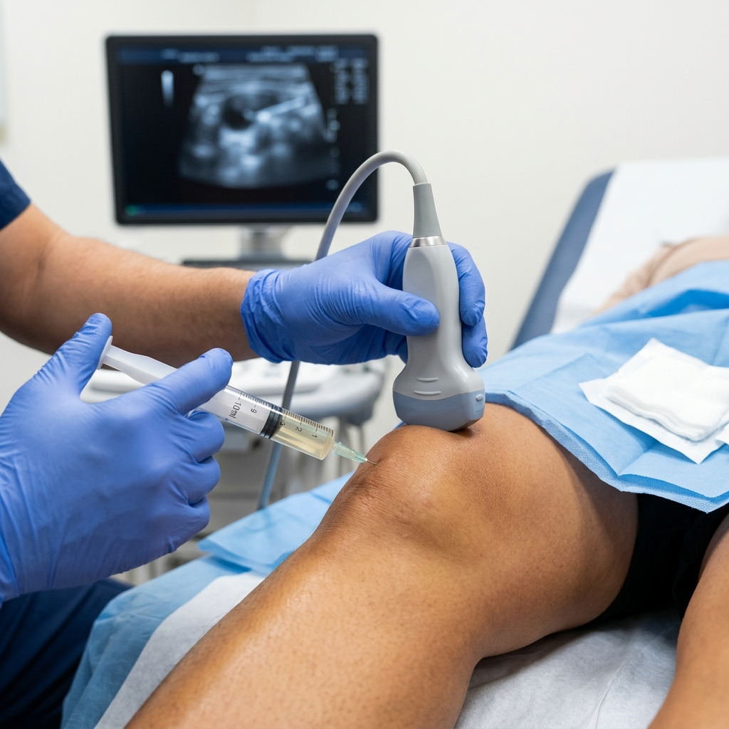
To stop the pain, the fluid must be physically removed. Therapeutic Arthrocentesis (joint aspiration) is the medical equivalent of opening a pressure valve. Dr. Rabara utilizes live diagnostic ultrasound to pinpoint the exact deepest pocket of fluid. We guide a small needle safely into the joint and gently suction the fluid out. The moment the fluid is extracted, the mechanical pressure drops to zero, and the ability to bend your joint is instantly restored.
Joint Swelling We Actively Drain
Do You Need Your Joint Drained Today?
Recognizing the Hydraulic Crisis
An acute effusion is not a subtle injury. It is a physical, visible crisis that severely limits your mobility and demands immediate medical attention.
Ideal indications
Knee, elbow, or ankle is visibly swollen to nearly twice its normal size.
Tight, throbbing pressure that gets worse when you try to bend the joint.
Massive, tight bulge behind your knee (Baker's Cyst) that makes straightening leg impossible.
Diagnosed history of Gout and experiencing a sudden, blindingly painful attack.
Clinical precautions
Severe, active skin infection (cellulitis) directly over the injection site.
Open, draining wound that could push bacteria into the joint space.
The Realities of Joint Draining
We understand the fear of having a needle inserted into an already agonizing joint. We want you completely prepared for the "Drain and Treat" process:
The "Two-Step" Protocol
Immediately after draining the fluid, we use the same needle to inject specialized medication to shrink the joint lining and "seal" the leak.
Immediate Relief, Temporary Weakness
The joint capsule has been stretched like an old rubber band. You must wear a compression wrap and rest for a few days to allow tissue to tighten back up.
Minimal Discomfort
We use powerful local anesthetic. Because we use ultrasound guidance, we go straight into the empty pocket without painfully scraping bone or cartilage.
Why You Cannot Drain a Joint "Blindly"
"Wait and See"
Applying ice and waiting for swelling to go down on its own. Inflammatory fluid acts like acid, destroying healthy cartilage if left to sit.
Ultrasound-Guided Arthrocentesis
Dr. Rabara sees the exact size, depth, and location of the fluid pocket on screen. Guaranteed 100% empty and accurate medicine delivery.
"Blind" Joint Aspiration
A doctor feels the outside of the joint and pokes based on landmarks. Frequently hits bone or misses deep pockets entirely.
What Happens After the Fluid is Gone?
The Compression Phase
You must wear a tight compression bandage (Ace wrap). This physically prevents the empty joint space from filling back up while medication works.
The Calming Phase
Anti-inflammatory medication takes full effect, shutting down the 'fire' inside. The joint may feel 'loose' as ligaments were stretched by swelling.
Addressing the Root Cause
We investigate the cause (meniscus tear, gout, arthritis) and prescribe Physical Therapy to ensure the swelling never returns.
Clinical Science & Technical Details
For our medical colleagues and highly analytical patients, we provide these transparent technical details on the pathophysiology and interventional protocols.
Explore the Clinical Science: Synovial Capsular Distension and Inflammatory Exudate
Normal synovial fluid is small in volume. During an acute inflammatory cascade, the synovial membrane dramatically increases vascular permeability, flooding the joint cavity with low-viscosity exudate rich in pro-inflammatory cytokines and degradative enzymes.
Rapid accumulation causes severe capsular distension, creating excruciating ischemic and mechanical pain. Evacuating this fluid is critical; if left to sit, degradative enzymes will actively destroy healthy articular cartilage.
Detailed Diagnostic Pathways: Synovial Fluid Analysis & Septic Arthritis
A clear, yellowish fluid typically indicates non-inflammatory osteoarthritis. Cloudy, watery fluid indicates a severe inflammatory process like Gout or Pseudogout.
Crucially, grossly purulent fluid suggests Septic Arthritis (bacterial infection). This is an absolute orthopedic emergency, and diagnostic aspiration allows for immediate fluid culture.
Advanced Systemic Screening and Baker's Cyst Rupture
For posterior knee swelling, Dr. Rabara must differentiate between a ruptured Baker's Cyst and Deep Vein Thrombosis (DVT) using diagnostic ultrasound.
If a cyst is intact but causing severe mechanical block, we perform ultrasound-guided aspiration frequently followed by fenestration of the cyst wall to prevent immediately re-accumulation.
Preventing Iatrogenic Infection and Rebound Effusions
While rare, Dr. Rabara utilizes rigorous aseptic technique. The skin is aggressively prepped with Chlorhexidine, and sterile ultrasound probe covers are mandated.
To prevent 'rebound effusion', we introduce a highly localized, long-acting corticosteroid into the newly evacuated space, suppressing the vascular permeability that caused the exudate.
Inside the Procedure: Suprapatellar Pouch Navigation and Dynamic Compression
Using a lateral approach with a linear transducer, Dr. Rabara visualizes the anechoic fluid collection in the suprapatellar recess, superior to the patella and deep to the quad tendon.
To ensure 100% evacuation, we apply dynamic manual compression ('milking') to the medial and lateral gutters, forcing loculated fluid into the suprapatellar pouch for extraction.
Restoring Capsular Integrity and Proprioceptive Function
A massive joint effusion causes Arthrogenic Muscle Inhibition (AMI)—a reflexive neurological shut-down of the VMO muscle to protect the joint.
Post-aspiration rehab is not optional. Dr. Rabara refers patients to our therapy team to re-establish neuromuscular control and proprioceptive balance training.
Comprehensive Joint Drainage & Aspiration Guides
Expert resources for 'water on the knee', gout, and Baker's cysts.
Water on the Knee
Symptoms, causes, and why tight joint swelling demands rapid release.
Gout & RA Swelling
Managing severe inflammatory attacks and hydraulic joint pressure.
Home Remedies vs. Draining
Why ice packs fail for 'over-inflated' joints and when to call a doctor.
The Aspiration Procedure
Step-by-step: How we safely drain fluid to provide instant pain relief.
Ultrasound Accuracy
Why diagnostic ultrasound is mandatory for safe, painless joint drainage.
Knee Aspiration Cost
2026 pricing and PhilHealth info for joint drainage in Vigan City.
Tubig sa Tuhod
Bakit namamaga ang tuhod? Sintomas at lunas para sa tubig sa joints.
Gamot sa Pamamaga
Paano alisin ang tubig sa tuhod nang mabilis gamit ang iniksyon (Tagalog).
Magkano Magpatanggal?
Mabilis at affordable na pagtanggal ng tubig sa tuhod sa Vigan clinic.
The Joint Drain and Treat Protocol
The Ultrasound Target Mapping
Dr. Rabara uses high-resolution ultrasound to map a needle trajectory that completely avoids your bones, cartilage, and major blood vessels.
The Pressure Release
After numbing the skin, we gently suction the inflammatory fluid while watching the screen. You will feel agonizing, tight pressure disappear in seconds.
The "Sealant" Injection
We leave the needle in place and inject targeted medication (Steroid or Gel). This calms the joint lining, ensuring the swelling does not return tomorrow.