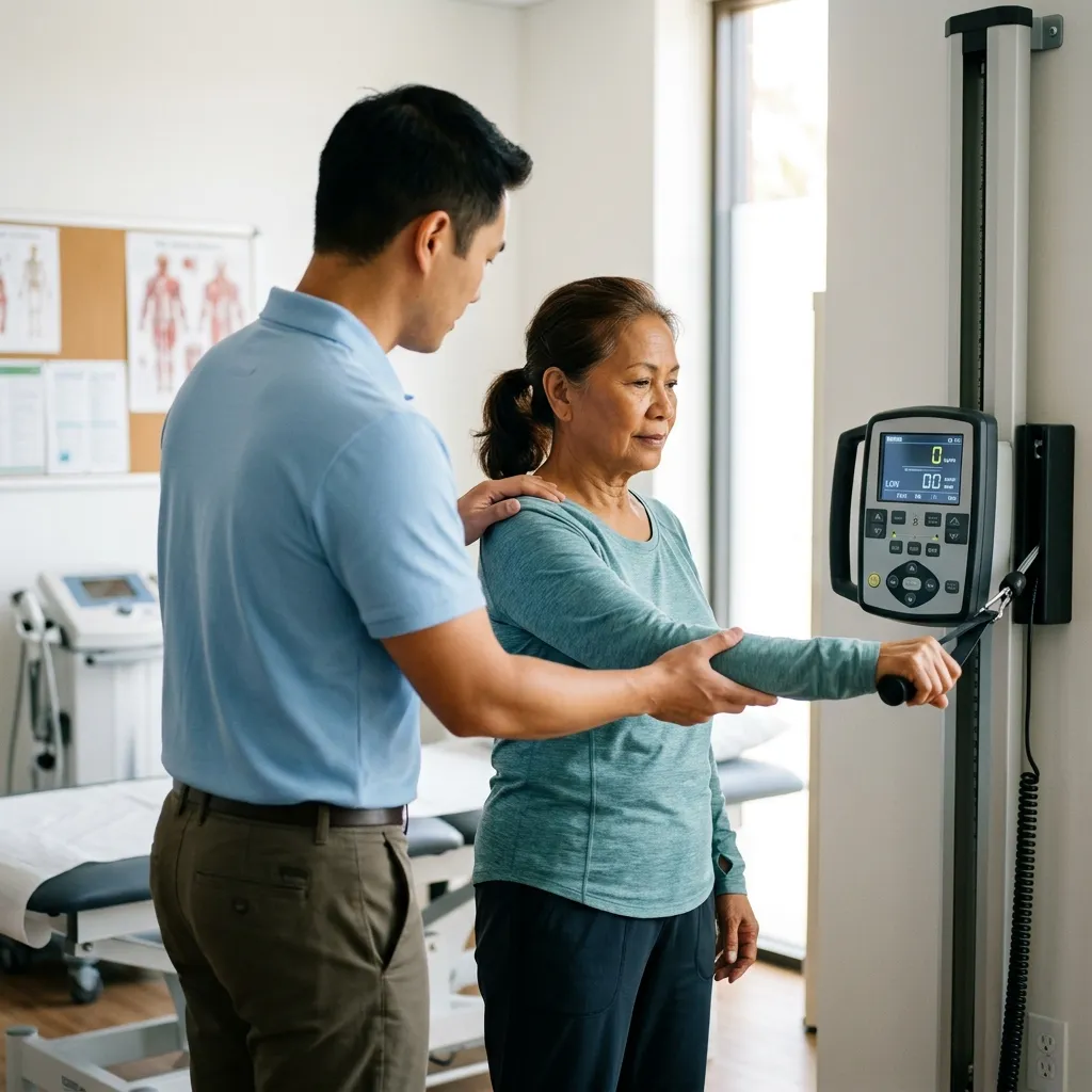
- — A bone density test measures bone mineral density at selected sites. It is interpreted with your age, sex, fracture history, medical risks, medicines and other clinical information.
- — A DEXA or DXA scan is quick and non-invasive, but the number is not a complete personal forecast and is not the same as an X-ray for a suspected acute fracture.
- — Osteopenia and osteoporosis describe different ranges of bone density, but fracture risk is not determined by one number alone.
- — If a report is confusing, ask for a clinical explanation rather than starting supplements, stopping activity or deciding that you are “fine” from the label alone.
A bone density test is an examination that estimates the mineral content of selected bones. It can be confusing because the report may show several sites, more than one score and labels such as osteopenia or osteoporosis. A DEXA scan is useful, but it is not a simple “good” or “bad” verdict. The result needs to be read with your age, fracture history, falls, medicines, nutrition, other health conditions and the reason the test was ordered.
For the nutrition and activity side of bone health, see our companion guide: How to Make Bones Stronger: Calcium, Vitamin D & Osteoporosis Diet.
In my clinic, I ask what the person is feeling as well as what the report says: a sharp or dull back pain, a stiff change in movement, radiating pain, tingling, burning, catching or grinding after a fall. Those symptoms do not diagnose osteoporosis, but they can change whether the next step is a routine explanation or prompt examination.
What does a bone density test measure?
A bone density test estimates bone mineral density, usually at selected areas such as the hip and lower spine. A DEXA or DXA scan is a common method. It helps a clinician classify bone density, assess fracture risk and establish a baseline when monitoring treatment; it does not measure pain, muscle strength, balance or every cause of a fracture (Feingold et al., 2000; PMID: 25905277).
The report is one piece of an assessment. A clinical evaluation may also include a medical history, physical examination, laboratory tests and, when indicated, other imaging. This matters because low bone mass can be related to nutrition, medicines, endocrine conditions, kidney problems, falls or another skeletal disorder. A number without that context can lead to either unnecessary fear or false reassurance.
What is a dexa scan and how does it work?
A DEXA scan uses low-dose X-rays to estimate bone mineral density at selected skeletal sites. You normally lie still while the scanner passes over the area being measured. The scan is not the same as an emergency X-ray for a new injury, and it does not show every detail of bone structure or tell you why a particular area hurts.
Before the appointment, tell the imaging team if you could be pregnant, recently had a contrast study, cannot lie flat, or have difficulty transferring. Follow the facility’s instructions. Ask which sites will be measured and when the report will be explained. The test is most useful when the result can be connected to a clear clinical question.
Bone mineral density measurement is widely used to detect, classify and monitor osteoporosis, while more specialized bone histomorphometry is largely used in research and academic settings (Bouxsein et al., 2018; PMID: 29955631). That distinction helps explain why a routine DEXA is often the first test discussed, but also why it cannot answer every bone-health question.
What does low bone density mean?
Low bone density means the measured mineral density is below the reference range used in the report. It does not automatically mean that you will have a fracture, that your bones are painful, or that one supplement will correct the problem. The result should prompt a risk conversation, not a self-diagnosis.
Osteopenia generally describes a lower-than-expected range that is not as low as the range used for osteoporosis. Osteoporosis is a more severe loss of bone density and strength, but the label is not the only factor that matters. A person with osteopenia may still have a meaningful fracture risk, while a person with a single borderline number may need the rest of the report and clinical history reviewed.
Previous fragility fracture, repeated falls, frailty, medicines that affect bone, low energy availability, menopause-related changes and other medical conditions can change the plan. The evaluation of a person at high fracture risk should look for factors contributing to low bone mass, skeletal fragility, falls and fractures (Feingold et al., 2000; PMID: 25905277).
“What does a T score mean” when reading a result?
A T-score compares a person’s bone mineral density with a young-adult reference population and expresses the difference in standard-deviation units. In general, a more negative number means a lower density compared with that reference. A commonly used threshold for osteoporosis in appropriate populations is at or below −2.5, but a clinician must confirm that the score, reference population, skeletal site and patient age are appropriate.
Do not decide that the lowest number is the entire diagnosis without asking which site it came from and whether it is technically reliable. Spine and hip results can differ. Degenerative change, positioning, hardware, scoliosis, a previous fracture or another technical issue can affect interpretation. Reports of unexpected bone-mass values can also have artefactual causes, which is why a clinician may review images and technical notes rather than reacting to a single number (Nottez et al., 2020; PMID: 32534222).
A T-score is not the same as a Z-score. A Z-score compares you with people of a similar age and sex, and it may be more relevant in some younger patients or specific clinical situations. Ask the clinician who ordered the test which score should guide the next decision.
“How bone density test works”: what happens during the scan?
The practical process is usually simple: confirm the reason for testing, review relevant history, position you on the scanner, acquire measurements at selected sites, and have the report interpreted. The scan itself does not tell you whether your diet is adequate, whether a medicine is affecting your bones, or whether a movement is safe after a fracture.
Bring the name and dose of medicines and supplements, previous scan reports if available, details of any fracture after a minor fall, recent height change, falls, and conditions such as kidney disease, thyroid or parathyroid disease, an eating disorder or early menopause. These details can be more useful than memorizing one score.
Do not compare two reports as if they were identical photographs. The scanner, facility, body position, measured site, reference database and time between tests matter. A clinician may also consider whether the change is larger than the facility’s measurement error before calling it a true change.
| Report item | What it helps describe | What it does not answer alone |
|---|---|---|
| Measured site | Where bone mineral density was estimated | Whether every bone or every pain source is normal |
| T-score or Z-score | How the result compares with a reference group | Your complete fracture risk or treatment need |
| Technical comment | Whether positioning, hardware or artefact may affect reading | Whether a new injury is safe to ignore |
| Clinical plan | What to do next based on the whole assessment | A guarantee that the score will never change |
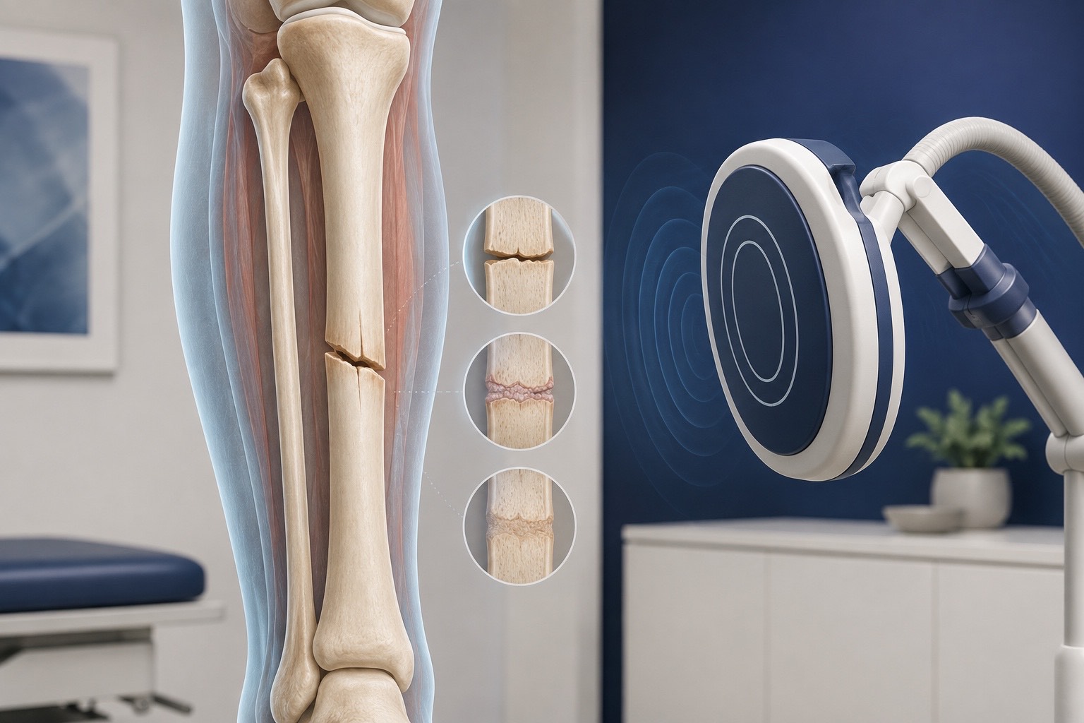
“When to get a bone density test”: who should ask?
Ask a clinician about testing after a low-trauma fracture, when a medicine or medical condition may weaken bone, when there is unexplained height loss or suspected vertebral fracture, or when your overall fracture risk makes a baseline useful. Screening recommendations vary by age, sex, country and risk factors, so a general internet rule cannot replace a clinical decision.
Testing may be especially important when the result could change management. A clinician may combine the scan with fracture-risk tools, examination, laboratory studies or imaging. The point is not to order a scan for every worry; it is to use the right test for the right question and act on the answer.
Access and cost are real concerns. Fees, equipment and coverage differ by facility and location, and this educational page does not verify a current Philippine price or PhilHealth benefit. Call the imaging facility or your insurer for current details, and ask your clinician whether the test is medically indicated before paying for it.
What should I do after a bone density result?
- Ask which site and score matter. Write down the hip, spine or other site, the score type, and any technical comment.
- Ask how your history changes the result. Mention fractures, falls, medicines, menopause, nutrition, kidney health and family history.
- Ask what action is needed now. The next step may be nutrition, exercise, fall prevention, medication discussion, laboratory review, another scan later, or no immediate change.
- Ask what symptoms need review. New back pain, loss of height, hip or groin pain, or a fall may need assessment even if the report label sounds mild.
Patient understanding is part of safety. Osteoporosis health literacy can be limited, and experience with a bone density test is associated with understanding risk in some populations (Lin et al., 2020; PMID: 32646011). It is reasonable to ask the clinician to explain the report in plain language and to write down the plan.
Can you exercise after a bone density test? The scan itself does not decide whether an activity is safe. Your movement plan should reflect pain, fracture history, balance, strength, posture and the clinician’s interpretation. Walking, resistance exercise, balance training and other activities may be useful for some people, while high-impact work, loaded spinal flexion or twisting may need modification after particular fractures or symptoms.
Do not stop all movement because a report contains the word osteopenia, and do not start jumping or heavy lifting solely because an online post recommends it. A rehabilitation assessment can identify a safe starting point, teach technique and set a progression that fits your real daily life.
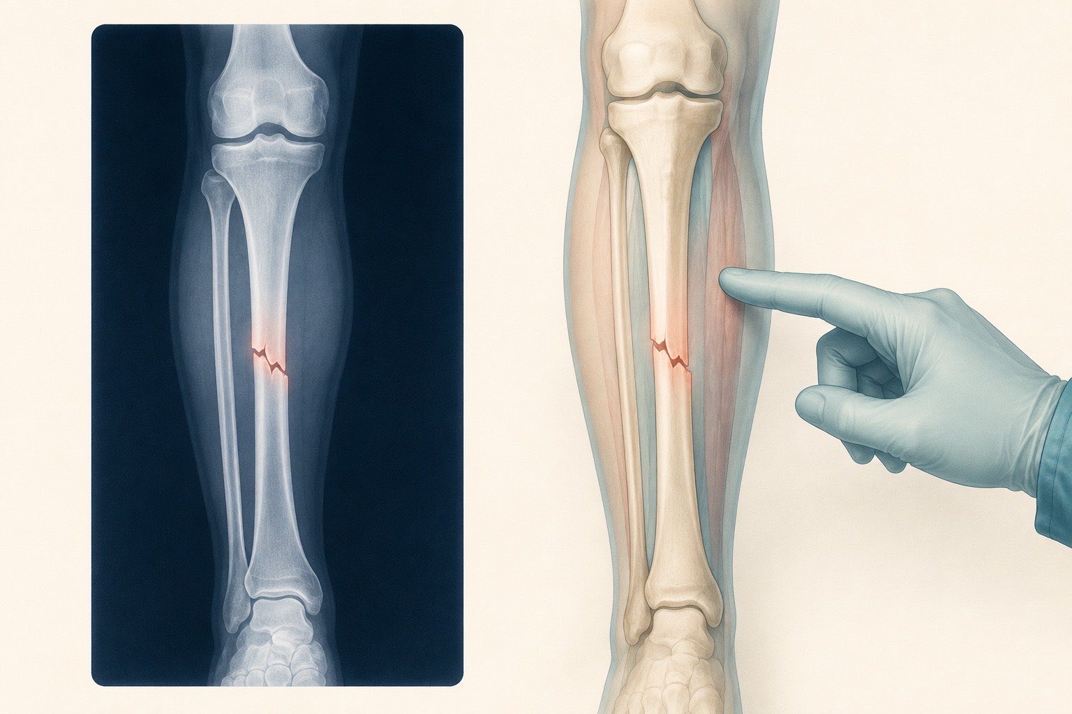
Need help understanding a report?
A Vigan rehabilitation assessment can translate the result into practical questions about fracture risk, movement, falls and next steps.
When should I seek urgent help? Seek urgent medical care after a major fall, sudden severe back or hip pain, inability to stand or bear weight, new weakness or numbness, or a limb that appears shortened or deformed. A routine bone density appointment is not a substitute for emergency assessment of a possible fracture.
A scan is a starting point for a conversation
The useful result is an understandable plan, not a frightening number without context.
Read the whole report
Sites, reference population, score type and technical comments can change how a result should be understood.
Add clinical risk
Previous fractures, falls, medicines, nutrition, menopause and other conditions may change the decision beyond the score.
Choose the next step
The next step may be reassurance, prevention, repeat testing, laboratory review, rehabilitation or medication discussion.
References & Clinical Evidence
- [1] Feingold KR, Adler RA, Ahmed SF, et al. Osteoporosis: Clinical Evaluation. Endotext. Updated 2000. PMID: 25905277. https://pubmed.ncbi.nlm.nih.gov/25905277/
- [2] Nottez A, Kolta S, Lion G, et al. Prevalence and causes of elevated bone mass. Bone. 2020;134:115476. PMID: 32534222. https://pubmed.ncbi.nlm.nih.gov/32534222/
- [3] Bouxsein ML, et al. Bone histomorphometry in the evaluation of osteomalacia. Bone Rep. 2018;9:155-161. PMID: 29955631. https://pubmed.ncbi.nlm.nih.gov/29955631/
- [4] Papaioannou A, et al. Screening, prevention and management of osteoporosis among Canadian adults. Health Promot Chronic Dis Prev Can. 2017;37(12):453-462. PMID: 30540411. https://pubmed.ncbi.nlm.nih.gov/30540411/
- [5] Lin LP, Lai WJ, Hsu SW, Lin JD. Health Literacy of Osteoporosis Risks among Caregivers Serving in Disability Care Facilities. Int J Environ Res Public Health. 2020;17(13):4903. PMID: 32646011. https://pubmed.ncbi.nlm.nih.gov/32646011/
- [6] Chen G, Liu H, Wu X, et al. Biomarkers of postmenopausal osteoporosis and interventive mechanism of catgut embedding in acupoints. Medicine (Baltimore). 2020. PMID: 32925788. https://pubmed.ncbi.nlm.nih.gov/32925788/
* Clinical references are provided to support the medical claims made in this article. TeraCare adheres to evidence-based practices in physical medicine and rehabilitation.

Dr. Ben Rabara
Dr. Ben Rabara is a Board-Certified Physiatrist specializing in Physical Medicine and Rehabilitation. He focuses on non-surgical, precision treatments for musculoskeletal conditions, utilizing advanced diagnostics like MSK Ultrasound.
Medical Disclaimer: The information provided in this article is for educational purposes only and does not substitute for professional medical advice, diagnosis, or treatment. Always consult a qualified physician for your specific health conditions.