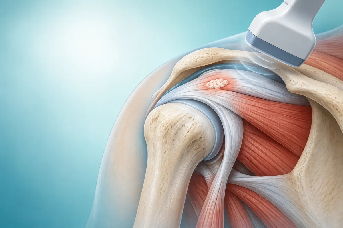
- — A calcium deposit in the shoulder cannot be safely dissolved at home. The right plan depends on the diagnosis, symptoms, imaging, deposit features, health history, and response to earlier care.
- — Calcific tendonitis can settle over time for some people, but severe pain or restricted movement may need a structured review of observation, rehabilitation, medication, injections, shockwave therapy, ultrasound-guided barbotage, or surgery.
- — Ultrasound-guided barbotage uses imaging to guide a needle toward a suitable deposit while fluid and gentle needling are used to break up or wash out some of the calcium. It is not appropriate for every shoulder or every deposit.
- — Do not inject, puncture, scrape, or take “calcium dissolving” supplements for shoulder pain without a clinician’s assessment. Bring your scan reports and medicine list to the consultation.
A calcium deposit in the shoulder is not something to dissolve at home. Calcific tendonitis can improve over time for some people, but persistent severe pain or restricted movement may lead a clinician to discuss observation, rehabilitation, injections, shockwave therapy, ultrasound-guided barbotage, or surgery. The safest option depends on the diagnosis, the deposit’s appearance and location, other shoulder findings, your health history, and how you respond to earlier care.
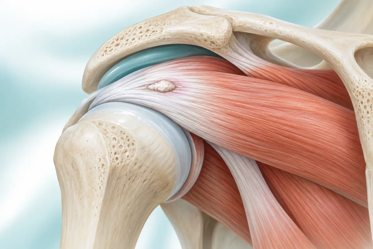
The word “calcium” can make this sound like a nutrition problem. In calcific tendonitis, calcium-containing material is found in or near a tendon, often in the rotator cuff. It is a local shoulder condition, not proof that you have too much calcium in your diet. A scan finding still needs to be matched with your symptoms and examination because a deposit may be present without being the only reason a shoulder hurts (Louwerens et al.; PMID: 37819397).
What is calcific tendonitis, and why can it hurt so much?
Calcific tendonitis is a painful shoulder condition involving calcium deposits in a tendon. The rotator cuff is a group of tendons that helps centre and move the shoulder. When a deposit irritates the tendon or nearby bursa, a person may feel pain with lifting, reaching behind the back, lying on that side, or moving through a particular part of the range. Some episodes are sudden and intense, with marked stiffness and difficulty using the arm.
The course is not identical for everyone. A deposit may be relatively quiet, become painful during an inflammatory phase, or remain associated with symptoms after the acute flare settles. A visible deposit does not automatically explain every symptom. Neck pain, adhesive capsulitis, rotator-cuff tearing, arthritis, nerve irritation, and referred pain can overlap with a “calcium deposit in shoulder” story and may change the treatment plan.
Assessment usually combines a history, shoulder and neck examination, and imaging chosen for the clinical question. X-ray may show calcific material; ultrasound can assess the deposit, tendon, bursa, and movement in real time; and other imaging may be considered when a different problem is suspected. The useful question is not only “How large is the calcium?” but also “Does this finding fit the pain and loss of function?”
What does a calcium deposit in shoulder mean?
“Calcium deposit in shoulder” is a plain-language description, not a complete diagnosis. The deposit may be within a tendon, close to the tendon, or in a location where it irritates nearby tissue. Its texture, stage, size, and relationship to the tendon can affect whether an image-guided procedure is technically reasonable. A report that says “calcification” should therefore be reviewed with the images and examination rather than treated as a stand-alone answer.
It is also possible to have more than one shoulder issue. A painful shoulder can have a deposit plus bursitis, tendon degeneration, weakness, or stiffness. If the pain is mainly in the neck, travels below the elbow, includes tingling, or follows an injury, the clinician may consider other causes as well. This is one reason a treatment chosen from a search result may be a poor fit for an individual patient.
Bring the original scan or a clear report, the date symptoms started, activities that make the pain worse, your current medicines, and any previous injections or therapy. These details help distinguish a new acute flare from a persistent mechanical or neurological problem and make the consultation more efficient.
How can you dissolve calcium deposits in shoulder safely?
There is no safe home technique that reliably dissolves a shoulder calcium deposit. Do not puncture the skin, inject a product bought online, massage aggressively over a hot painful area, or take a “calcium dissolving” supplement without medical advice. These approaches can cause infection, bleeding, medication interactions, delayed diagnosis, or more irritation while leaving the actual problem untreated.
A clinician may first recommend a period of observation, activity modification, pain relief that is safe for your medical history, and rehabilitation. Some deposits change over time without an invasive procedure, while others remain painful or continue to restrict function. The decision to wait should have a review point and a plan for what to do if pain, weakness, or motion worsens.
If symptoms remain limiting, treatment may be discussed in stages. Options can include a clinician-guided injection for selected pain generators, shockwave therapy, ultrasound-guided barbotage, or referral for surgery in a smaller group of patients. Evidence differs between treatments and studies; an option being available does not mean it is appropriate for every deposit or guaranteed to remove pain (Ibrahim et al.; PMID: 36684664).
What is ultrasound guided barbotage shoulder treatment?
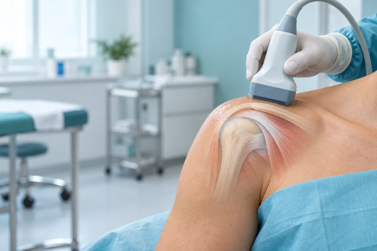
Ultrasound-guided barbotage is a procedure intended for selected calcific deposits. The clinician uses ultrasound to identify the tendon, deposit, nearby bursa, and important structures. After discussing risks and comfort measures, a needle may be directed toward the deposit and fluid may be used with gentle needling to break up and aspirate some of the material. The exact technique varies between clinicians and deposits (Sconfienza et al.; PMID: 34283462).
The ultrasound component is important because it helps the clinician see the target and avoid structures that should not be traversed. It does not make the procedure risk-free, and it does not mean that every visible deposit can be reached or removed. A suitable plan should include the working diagnosis, expected benefit, alternatives, possible complications, aftercare, and what happens if the shoulder remains painful.
You may feel pressure, brief needle discomfort, or soreness afterwards. Ask before the procedure how the area will be numbed, whether fluid will be placed in a bursa, what movements are allowed afterwards, and which symptoms should prompt a call. Image guidance can improve targeting, but it cannot replace sterile technique, clinical judgement, or a rehabilitation plan.
Who might be considered for barbotage calcific tendinitis?
Barbotage may be considered when the symptoms and imaging fit calcific tendinitis, the deposit is technically approachable, and non-procedural care has not restored useful function. The decision is individual. A large or easily seen deposit is not by itself an indication, and a small deposit can still be painful for reasons that barbotage will not address.
A clinician may pause or choose another approach when there is uncertainty about the diagnosis, active infection, an important bleeding risk, an unsafe medication issue, a suspected major tear, or a pain pattern suggesting the neck or nerves. Tell the clinician about blood thinners, allergies, diabetes, immune problems, pregnancy, prior shoulder surgery, and any recent fever or skin infection.
Ask whether the proposed procedure is meant to reduce pain, improve movement, remove some deposit, or test a pain source. These are different goals. Clear goals make it easier to judge progress and avoid interpreting a temporary change as proof that every underlying problem has been solved.
What is calcific tendinitis treatment without surgery?
Non-surgical care usually starts with a plan to settle pain while protecting, but not completely immobilising, the shoulder. The clinician may recommend short-term activity changes, a suitable analgesic or anti-inflammatory approach, and exercises that maintain comfortable movement. Medication advice must account for kidney disease, stomach or heart risk, pregnancy, anticoagulants, allergies, and other medicines.
Rehabilitation is not a punishment for having a deposit. It is a way to restore usable movement, strength, and confidence after the acute pain is understood. A therapist may begin with comfortable range-of-motion work, then progress to rotator-cuff and shoulder-blade strengthening as tolerated. The exact sequence should be adjusted if the shoulder is highly irritable, very stiff, weak, or recently treated.
Shockwave therapy, injections, and other options may be discussed when symptoms persist. Systematic reviews and clinical studies do not mean that one modality is automatically best for all patients. Ask what evidence applies to your deposit, how many sessions are proposed, how benefit will be measured, and what the next step will be if the plan does not help.
How does barbotage calcific tendinitis care compare with other options?
A useful comparison focuses on the problem being treated, the expected time course, and the uncertainty. Observation may avoid a procedure but requires a safety net. Rehabilitation can improve function but may be difficult during a severe flare. An injection may target inflammation or pain but does not necessarily remove a deposit. Shockwave therapy may be considered for selected chronic cases, while barbotage aims to address suitable calcium under imaging. Surgery is generally reserved for carefully selected persistent cases after discussion of alternatives.
| Option | What it may aim to do | Important question |
|---|---|---|
| Observation and rehabilitation | Settle symptoms and restore movement over time | When will progress be reviewed? |
| Shockwave or injection | Address selected pain or tendon-related problems | What evidence fits this deposit and goal? |
| Ultrasound-guided barbotage | Break up or aspirate some suitable calcium | Is the deposit technically appropriate and what is the fallback plan? |
| Surgery | Address selected persistent structural problems | Have diagnosis and non-surgical options been reviewed? |
The safest choice is the one that matches the confirmed problem and your priorities. If a clinician recommends a procedure, ask about the person’s experience with the technique, sterile precautions, likely soreness, signs of infection, movement restrictions, rehabilitation, and follow-up. If the plan changes because the deposit is not suitable, that is a reason to reassess—not a reason to chase a more aggressive treatment.
What can recovery involve after calcific tendonitis treatment?
Recovery is measured by function as well as pain. You may be asked to keep the shoulder moving within a comfortable range, avoid heavy or repetitive loading for a short period, and then progress exercises gradually. Some people improve quickly; others need more time, especially when the shoulder has been stiff or guarded for weeks.
Follow the specific aftercare instructions given for your procedure. Do not add extra exercises because a video promises a faster result, and do not stop using the arm completely unless instructed. Contact the treating team for increasing redness, warmth, drainage, fever, severe swelling, new weakness, progressive numbness, or pain that is rapidly worsening rather than following the expected course.
When should you seek reassessment for shoulder pain?
Arrange a clinical review when shoulder pain keeps limiting sleep, work, dressing, reaching, or exercise; when movement is becoming more restricted; when weakness is new or progressive; or when symptoms continue despite a reasonable initial plan. Ask for the diagnosis to be revisited if the scan finding and pain pattern do not match or if treatment has not produced the expected change.
Seek urgent assessment for a shoulder injury with deformity or inability to use the arm, a hot swollen joint or fever, rapidly spreading redness, severe unrelenting pain, new numbness or marked weakness, chest pain, shortness of breath, or symptoms that make you acutely unwell. These warning signs should not be attributed to calcium without an examination.
If you are considering care in Vigan or nearby, bring your original images or report, medicine list, allergies, relevant medical conditions, and a brief timeline of the pain. Ask which options are available, whether the clinician performs the procedure, how suitability is decided, and what follow-up would be provided. Availability and fees change, so confirm them directly rather than relying on an old page or a social-media post.
Calcific tendonitis is treatable, but it is not a problem that can be safely solved by a universal home remedy. A careful diagnosis, realistic discussion of options, and graded recovery plan are more valuable than a promise that every deposit can be dissolved in one session.
Read our diagnostic musculoskeletal ultrasound guide to understand how imaging may fit into a broader assessment.
Calcific Tendonitis Treatment Realities
A shoulder calcium deposit is a finding that needs clinical context, not a promise of instant removal.
The deposit is not always the whole problem
Pain and loss of movement can also involve bursitis, rotator-cuff disease, adhesive capsulitis, arthritis, nerve irritation, or another condition. Imaging and examination need to be considered together.
Treatment response varies
The size, consistency, location, and stage of a deposit may affect which options are reasonable. Improvement may be gradual, and an intervention may not remove every source of pain.
Rehabilitation still matters
After pain is controlled, a carefully graded movement and strengthening plan can help restore function. The exercises and pace should match the examination and any procedure instructions.
References & Clinical Evidence
- [1] Louwerens JKG, et al. Calcific tendinitis of the shoulder: a review. 2021. PMID: 37819397.
- [2] Sconfienza LM, et al. Ultrasound-guided barbotage for calcific tendinitis of the shoulder. 2018. PMID: 34283462.
- [3] McCulloch PC, et al. Ultrasound-guided musculoskeletal injections. 2021. PMID: 34937117.
- [4] Oliva F, et al. Calcific tendinopathy of the rotator cuff: pathogenesis, diagnosis and treatment. 2015. PMID: 29946204.
- [5] Ibrahim MI, et al. Extracorporeal shock wave therapy for chronic calcific tendonitis of the shoulder: systematic review. 2022. PMID: 36684664.
- [6] Khan KM, et al. Physical therapy and exercise in the management of shoulder calcific tendinopathy. 2018. PMID: 29963844.
* Clinical references are provided to support the medical claims made in this article. TeraCare adheres to evidence-based practices in physical medicine and rehabilitation.

Dr. Ben Rabara
Dr. Ben Rabara is a Board-Certified Physiatrist specializing in Physical Medicine and Rehabilitation. He focuses on non-surgical, precision treatments for musculoskeletal conditions, utilizing advanced diagnostics like MSK Ultrasound.
Medical Disclaimer: The information provided in this article is for educational purposes only and does not substitute for professional medical advice, diagnosis, or treatment. Always consult a qualified physician for your specific health conditions.