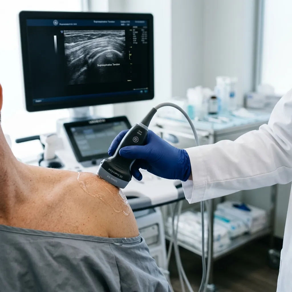
What is Diagnostic Musculoskeletal (MSK) Ultrasound?
For decades, patients with joint or tendon pain were sent for X-rays (which cannot see soft tissue) or expensive MRIs (which require lying perfectly still). Musculoskeletal (MSK) Ultrasound revolutionizes the diagnostic process. Utilizing high-frequency sound waves, this advanced imaging technology creates high-resolution, real-time pictures of your muscles, tendons, ligaments, nerves, and joints entirely without the use of harmful radiation.
The greatest clinical advantage of MSK Ultrasound is that it is a dynamic study. Pain rarely happens when you are lying perfectly still; it happens when you move. Unlike an MRI, an ultrasound allows Dr. Rabara to actively move your joint while scanning the tissue. We can actually watch the tendon glide, observe the ligament stretch, and pinpoint exactly when and where the impingement or inflammation occurs.
What Conditions Can MSK Ultrasound Detect?
Is MSK Ultrasound
the Right Test for You?
Musculoskeletal Sonography is the gold standard for dynamic tissue assessment.
Ideal indications
Suspected rotator cuff or Achilles tendon tears.
Lumps or bumps that need soft-tissue differentiation.
Nerve entrapments like Carpal Tunnel Syndrome.
Pain that only occurs during specific joint movements.
Clinical precautions
Suspected deep joint pathology (e.g., meniscus tears deep in the knee or spinal disc herniations).
Evaluation of internal bone structure or marrow abnormalities.
Diagnostic MSK Ultrasound Realities
Understanding the scope and limitations of diagnostic ultrasound.
Operator Dependency
The accuracy of ultrasound imaging relies heavily on the skill and specific training of the interpreting physician.
Depth Limitations
Ultrasound cannot penetrate bone, meaning it cannot visualize deep intra-articular structures (like the ACL or deep meniscus) as clearly as an MRI.
Dynamic vs. Static
While excellent for viewing moving structures (dynamic), it may not provide the comprehensive overview that a static MRI slice offers.
How Does Ultrasound
compare to Other Scans?
High-Resolution Soft Tissue Imaging Without Radiation
Advanced Scanning
Technology
What to Expect During Your Real-Time Scan
Real-Time Visualization: The Tech Behind the Probe
Musculoskeletal ultrasound relies on the principle of acoustic impedance to visualize tissue architecture. Normal tendons exhibit a highly organized, hyperechoic "fibrillar pattern" when viewed longitudinally—often described as a series of parallel linear echoes representing collagen fascicles.
A critical technical concept in MSK ultrasound is Anisotropy. Because of the linear arrangement of tendon fibers, if the ultrasound beam is not perfectly perpendicular to the tissue, the tendon will artifactually appear dark, potentially mimicking a tear. This is why specialized training in ultrasound physics and beam angle optimization is mandatory.
Dynamic Testing: The Biomechanical Advantage Over MRI
The single greatest clinical advantage of MSK Ultrasound is its dynamic capability. Traditional MRI and CT scans are "static"—they take a snapshot while the patient is perfectly still.
During your evaluation, Dr. Rabara can actively move your joint while the probe is on the skin, allowing us to watch the tendon glide beneath the bone in real-time. We can pinpoint exactly where the 'catch' or 'impingement' happens.
Diagnostic Advantages of High-Frequency Sound Waves
Unlike X-rays or CT scans, which utilize ionizing radiation, MSK ultrasound is based on the Piezoelectric Effect. The transducer probe contains specialized crystals that vibrate, producing sound waves.
Because there is no radiation, ultrasound is exceptionally safe for repeated use, monitoring healing progress over time, and for use in pediatric or pregnant patients.
The 3-Step Clinical Process
Targeted Physical Examination
We begin with a focused musculoskeletal exam to identify the specific area of pain and isolate the biomechanical dysfunction.
Real-Time Dynamic Scanning
Applying a water-based gel, Dr. Rabara glides the high-frequency probe over the affected area while you move the joint.
Immediate Diagnosis & Strategy
We explain the findings on the screen immediately and map out your recovery plan during the very same visit.
Clinical decision support
What musculoskeletal ultrasound can—and cannot—answer
Musculoskeletal ultrasound provides a real-time view of many superficial tendons, muscles, ligaments, bursae, joints, and peripheral nerves. Dynamic scanning can reproduce movement-related symptoms and comparison with the opposite side may add context. It does not replace every other test: deep structures, bone marrow, complex intra-articular disease, or findings that remain unclear may still require radiographs, MRI, laboratory testing, or referral.
A useful scan begins with a focused clinical question. An image finding is interpreted alongside the history and physical examination because structural changes can exist without causing pain. Urgent symptoms, suspected fracture, infection, tumor, or major neurologic loss are assessed through the appropriate emergency or specialist pathway rather than ultrasound alone.
How this evidence is applied at TeraCare
At TeraCare, Dr. Ben Rabara performs and interprets the scan in the context of the patient’s examination, explains whether a finding matches the symptoms, and documents when another test or referral would be more appropriate.
Medical review: Dr. Ben Rabara, Physiatrist · Evidence reviewed August 22, 2026. This information supports shared decision-making and does not replace an individual examination or emergency care.
Clinical sources: AIUM Practice Parameter for the Performance of a Musculoskeletal Ultrasound Examination (2023).
How this treatment fits your care plan
MSK, Joint & Tendon Care
Diagnosis, symptom control, tissue repair, and return to movement for joint, muscle, and tendon problems.
Your exact sequence depends on the diagnosis, safety screening, and response to treatment. A physiatrist will recommend only the steps that fit your case.
Where this treatment is available
Availability is generated from each active branch’s service record.
Common Questions
Is MSK Ultrasound safe for pregnant women or children?
Does the ultrasound scan hurt?
How long will the appointment take?
Why don't all doctors just use ultrasound instead of MRI?
Are the costs transparent for a cash-pay patient?
References & Clinical Evidence
- [1] Clinical guidelines and evidence-based research supporting Diagnostic MSK Ultrasound Imaging.
- [2] Relevant peer-reviewed literature on efficacy and safety outcomes in rehabilitation.
* Clinical references are provided to support the medical claims made in this article. TeraCare adheres to evidence-based practices in physical medicine and rehabilitation.

Dr. Ben Rabara
Dr. Ben Rabara is a Physiatrist in Vigan City specializing in Physical Medicine and Rehabilitation. He focuses on non-surgical, precision treatments for musculoskeletal conditions, utilizing advanced diagnostics like MSK Ultrasound.
Medical Disclaimer: The information provided in this article is for educational purposes only and does not substitute for professional medical advice, diagnosis, or treatment. Always consult a qualified physician for your specific health conditions.
MSK Ultrasound Diagnostic Guides
Explore how real-time, dynamic imaging pinpoint muscle, tendon, and joint injuries without radiation.