- — Tendonitis is a tension injury caused by pulling forces on muscle attachments; it requires progressive loading, not rest, to stimulate cellular repair.
- — Bursitis is a compression injury caused by friction on cushioning sacs; it requires unloading and pressure relief.
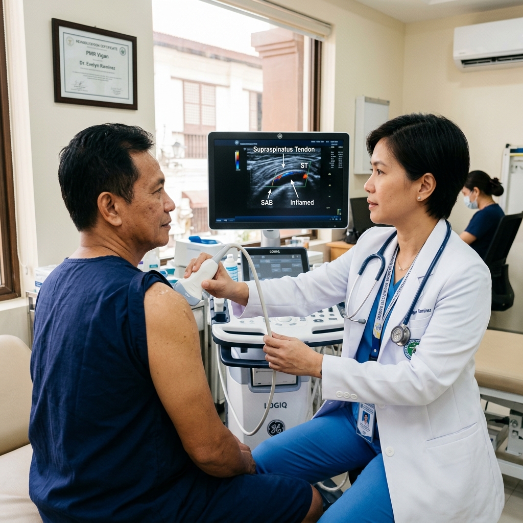
- — Bedside Musculoskeletal (MSK) Ultrasound allows the physician to dynamically view tissues in active motion, instantly distinguishing these adjacent pain drivers.
The Anatomy of Joint Pain: What is Tendonitis and Bursitis?
Tendonitis and bursitis are distinct inflammatory joint conditions that represent separate anatomical failures. Tendonitis is the structural degeneration and micro-tearing of a tendon under mechanical tension, whereas bursitis is the swelling of a fluid-filled synovial bursa sac under mechanical compression. Differentiating these tissues is critical because they require opposite loading and recovery strategies.
I frequently meet patients in my Vigan clinic who use the terms "tendonitis" and "bursitis" interchangeably to describe their joint pain. While both conditions cause localized pain, stiffness, and frustration, they represent entirely different anatomical failures. A tendon is a thick, cord-like band of fibrous tissue that anchors muscle to bone, designed to transmit high-tension forces. Tendonitis (or more accurately, tendinopathy) is the micro-tearing and structural breakdown of these fibers under repetitive pulling stress. A bursa, on the other hand, is a small, fluid-filled sac lined by a synovial membrane that acts as a low-friction cushion between bones, tendons, and muscles. Bursitis is the swelling and inflammation of this cushion due to excessive friction or compression. Differentiating between them is not just an academic exercise; it changes your entire rehabilitation strategy.
In my clinical practice, I emphasize that these two conditions react to mechanical forces in opposite ways. A tendon is a living structure that adapts to load. When it degenerates, it requires progressive mechanical loading to stimulate cellular repair and rebuild tissue strength. A bursa, however, is a simple friction pad. When it becomes inflamed and swollen, direct compression or rubbing will only worsen the swelling. Treating them the same way—usually with complete rest and oral painkillers—frequently leads to chronic stiffness or further joint damage. My primary diagnostic task is to identify which structure has failed so we can apply the correct mechanical solution.
Contrarian Insight: Complete bed rest or prolonged immobilization is toxic to a degenerating tendon. While resting an inflamed bursa helps quiet the swelling, resting a tendon causes the healthy collagen fibers to atrophy and disorganize. When you eventually return to activity, the weakened tendon is even less capable of handling tension, leading to a vicious cycle of re-injury.
What Generic Medical Guides Miss About This Condition
Standard health websites often prescribe identical "RICE" protocols (Rest, Ice, Compression, Elevation) for all joint pain. In my clinical experience, complete immobilization is counterproductive for degenerating tendons, which need guided tension to align collagen fibers. Furthermore, generic guides omit that hard, aggressive massage (often sought via traditional local hilot treatments) can rupture an inflamed bursa, spreading inflammatory fluid and worsening joint pain.
Tension vs. Compression: How the Pain Feels Different
Tendonitis and bursitis pain present with distinct sensory patterns reflecting their mechanical drivers. Tendonitis causes sharp, stabbing pain along the tendon pathway that flares under muscle contraction, often described as a "tight cord pulling." Bursitis causes a constant, deep throbbing sensation resembling internal "bruising from the inside" that worsens under direct pressure, such as sleeping on the joint.
In my clinical assessments, I teach patients to pay attention to the mechanical triggers of their pain, as these provide key clues to the underlying pathology. Tendonitis is fundamentally a tension problem. The pain is felt when the associated muscle contracts, pulling on the damaged fibers. Patients describe this sensation as a "tight cord pulling" or a "hot wire" running along the side of the joint. The pain presents as a "sharp stab" during movement—such as gripping a heavy coffee mug, twisting a screwdriver, or lifting a grocery bag—and typically eases the moment the muscle is relaxed.
Bursitis is fundamentally a compression problem. Because the bursa is a fluid-filled sac, it acts like a "balloon inside the joint." When it inflames, the sac swells and puts constant pressure on surrounding nerves. Patients describe this as a deep, throbbing ache that feels like "bruising from the inside." Unlike tendonitis, which quietens down at rest, bursitis pain is persistent and highly sensitive to direct pressure. If you have shoulder bursitis, sleeping on your affected side will frequently wake you with a throbbing, burning pain. Leaning your elbow on a table or kneeling on the floor can trigger immediate, intense tenderness because you are directly squeezing an inflamed bursa against the bone.
Tendon and Bursa Overlap: Shoulder, Elbow, Hip, and Knee
Tendon and bursa overlap occurs frequently in major joints where tendons run immediately adjacent to protective synovial cushions. Repetitive joint movements cause these structures to compress and rub against one another, frequently producing coexisting tendonitis and bursitis. The shoulder subacromial space, the lateral elbow, the greater trochanter of the hip, and the patellar region of the knee are the primary clinical sites for this overlap.
When I evaluate patients presenting with acute or chronic joint pain, I must account for the close anatomical proximity of tendons and bursae. Because these structures lie immediately adjacent to each other to facilitate smooth movement, they frequently flare up together. It is very common for a patient to suffer from concurrent tendinitis and bursitis, where a degenerating tendon rubs against its protective bursa, triggering secondary swelling in the fluid sac.
In my practice, I map these overlaps to four primary clinical presentations:
- The Shoulder: The supraspinatus tendon lies directly beneath the subacromial-subdeltoid bursa. Repetitive overhead lifting can cause the bursa to swell, compressing the rotator cuff tendon and mimicking a tear.
- The Elbow: Tennis elbow (lateral epicondylitis) involves micro-tears in the forearm extensor tendons. However, leaning directly on the elbow can irritate the olecranon bursa, which sits right over the bony tip.
- The Hip: Greater Trochanteric Pain Syndrome (GTPS) often involves both gluteus medius tendinopathy and trochanteric bursitis. Differentiating between them is crucial, as the gluteal tendon requires progressive loading while the trochanteric bursa requires decompression.
- The Knee: Patellar tendinopathy (jumper's knee) causes pain directly below the kneecap. It frequently coexists with deep infrapatellar bursitis, where the bursa behind the patellar tendon becomes swollen and pinched during deep squats.
The Dynamic Diagnostic Key: Musculoskeletal (MSK) Ultrasound
Bedside musculoskeletal (MSK) ultrasound is the definitive dynamic tool for distinguishing tendonitis from bursitis in real-time. By utilizing high-frequency sound waves during active joint movement, the physician visualizes whether pain is driven by tendon fiber tearing or by synovial fluid distension. This point-of-care assessment avoids the delay of static MRIs and the structural blind spots of traditional X-rays.
To make a definitive diagnosis, I perform point-of-care musculoskeletal (MSK) ultrasound in my clinic. Traditional physical examinations can localise tenderness, but they cannot look through the skin to verify the tissue state. X-rays are blind to soft tissues, and standard MRIs are static, capturing the joint only in a neutral, resting position. MSK ultrasound uses high-frequency sound waves to produce high-resolution, real-time images, allowing me to dynamically stress the joint and watch the tissues interact in active motion.
During a shoulder scan, for example, I can measure the thickness of the subacromial-subdeltoid bursa. Current clinical evidence suggests that a bursa thickness exceeding 3.0 millimeters (mm) is highly specific and sensitive for active subacromial bursitis, helping me differentiate it from a degenerative rotator cuff tendon (Schmidt A, et al., 2025; PMID: 40892101). In hip pain, I use high-frequency transducers to visualize whether the pain is driven by a thickened, inflamed trochanteric bursa or by micro-tearing and fiber disorganization at the gluteus medius tendon insertion (Hilligsøe L, et al., 2020; PMID: 32381380). This live visualization allows me to make an immediate, accurate diagnosis during your visit.
Treatment Pathways: Why "Just Rest" is a Healing Trap
Rehabilitation pathways for joint pain must target the specific tissue failure to ensure proper healing. Tendonitis requires a customized, progressive loading program to stimulate collagen alignment and restore tendon strength, whereas bursitis requires strict joint offloading and biomechanical decompression to allow the inflamed sac to settle. Traditional aggressive massage is contraindicated as direct pressure can rupture a swollen bursa.
I structure my rehabilitation programs according to the specific tissue that has failed. Because tendons and bursae respond to mechanical forces in opposite ways, a generic "one-size-fits-all" rest protocol will fail. If I diagnose you with chronic tendonitis, I will prescribe a progressive loading program. Tendons require guided tension to stimulate tenocyte activity, lay down new collagen, and align fibers along the axis of force (Kim Y, et al., 2022; PMID: 35919692). If I diagnose you with bursitis, my immediate focus is decompression—offloading the joint, reducing direct friction, and correcting the biomechanical faults that are pinching the bursa.
Clinical Warning: I strongly advise my patients against seeking aggressive massage or traditional hilot rubbing on an actively swollen joint or bursa. Hard, localized compression on an inflamed bursa can damage its delicate synovial lining or even rupture the sac, spreading inflammatory fluid into the surrounding muscle planes and triggering a severe, debilitating pain flare.
Evidence-to-Practice Rehabilitation Framework
Evidence says:
Systematic reviews and randomized controlled trials show that eccentric and heavy slow resistance (HSR) training protocols are superior to passive modalities for tendinopathy, while bursitis responds rapidly to localized offloading and targeted anti-inflammatory measures (Kumar R, et al., 2022; PMID: 35340491).
In practice:
For many of my patients in Vigan, Ilocos Sur, traveling multiple times a week to a central rehabilitation facility is highly impractical due to work schedules, transport limitations, and the costs of repeated supervised sessions.
Why I may adapt:
I often prescribe a structured, home-based progressive exercise plan consisting of just two or three high-yield movements (such as heel drops for Achilles tendonitis or isometric gluteal holds for hip pain). This ensures excellent patient compliance, reduces travel stress, and delivers the necessary mechanical load safely at home.
What I watch for:
I monitor the patient's pain boundary closely. During tendon loading, a mild discomfort of 3/10 on the pain scale is acceptable, provided it settles back to baseline within 24 hours. However, any sharp, pinching pain during exercise indicates bursa compression and requires an immediate reduction in range or load.
When I escalate:
If a patient presents with severe, non-resolving bursitis that fails to respond to 4 weeks of offloading, or if they have a high-grade tendon tear confirmed on ultrasound, I escalate care. Under direct, real-time ultrasound guidance, I can perform a precise needle aspiration to drain the bursa fluid or deliver a localized injection to quiet the inflammation, followed immediately by targeted physical therapy.
Irritability-Based Rehabilitation Progression
To help patients recover safely, I structure exercise progression based on tissue irritability levels rather than time alone:
| Irritability Stage | Clinical Presentation | Tendon Protocol | Bursa Protocol |
|---|---|---|---|
| High (Acute) | Pain at rest, severe pain with minimal movement, sleep disruption. | Isometric contractions (holding a position for 30–45 seconds under light load) to reduce pain. | Strict offloading, temporary joint protection, cryotherapy, and side-sleeping adjustments. |
| Moderate (Subacute) | Pain only during active movement; settles quickly once movement stops. | Isotonic exercises (concentric/eccentric movements like calf raises) with controlled speed. | Gentle active-assisted ROM, avoiding end-range pinching or direct compression. |
| Low (Chronic) | No pain at rest; mild ache only after high-load or repetitive tasks. | Heavy slow resistance (HSR) and plyometric loading to restore full mechanical capacity. | Dynamic joint stabilization, eccentric strengthening of stabilizing muscles. |
Summary Comparison Table
The bursitis vs. tendonitis comparison table summarizes the critical mechanical, diagnostic, and clinical distinctions between these joint conditions. It serves as an active reference guide to help patients understand their tissue-specific rehabilitation pathways, typical pain presentations, and dynamic musculoskeletal ultrasound findings.
| Feature | Tendonitis | Bursitis |
|---|---|---|
| Structure Involved | Tendon (fibrous cord connecting muscle to bone). | Bursa (fluid-filled synovial friction sac). |
| Primary Cause | Repetitive tension load, pulling strain, overuse. | Excessive friction, repetitive compression, direct trauma. |
| Pain Quality | Sharp, localized, "hot wire" or "tight pulling" sensation. | Dull throbbing, deep ache, "bruised from the inside." |
| Direct Pressure | Mild tenderness along the tendon line. | Severe tenderness directly over the bursa sac. |
| Night Pain | Minimal, unless the joint is held in a stretched position. | Severe, especially when lying directly on the affected joint. |
| Rehabilitation Target | Progressive loading (isometric → isotonic → heavy slow resistance). | Offloading, biomechanical correction, and decompression. |
| Dynamic Ultrasound view | Hypoechoic (dark) thickening, fiber disorganization, micro-tears. | Synovial fluid distension, thickened bursa walls (>3mm). |
References & Clinical Evidence
- [1] Schmidt A, et al. Differentiating polymyalgia rheumatica from degenerative rotator cuff tendinopathy using subacromial-subdeltoid bursa thickness. Rheumatology. 2025. PMID: 40892101. DOI: 10.1007/s00296-025-05971-8.
- [2] Hilligsøe L, et al. Ultrasonographic assessment of greater trochanteric pain syndrome (GTPS): tendinopathy vs. bursitis. Journal of Ultrasound. 2020. PMID: 32381380. DOI: 10.1007/s40477-020-00456-4.
- [3] Kim Y, et al. Ultrasound-guided diagnostic and therapeutic injections for lateral epicondylitis and lateral epicondylalgia: tendonitis vs. tearing. Journal of Clinical Ultrasound. 2022. PMID: 35919692. DOI: 10.1002/jcu.23275.
- [4] Kumar R, et al. Coexistence of patellar tendinopathy and deep infrapatellar bursitis: ultrasound-guided diagnostic injections and therapeutic outcomes. Skeletal Radiology. 2022. PMID: 35340491. DOI: 10.1007/s00256-022-04021-z.
- [5] Page P, et al. Dynamic ultrasound evaluation of the Achilles tendon and retrocalcaneal bursa in runners. Journal of Athletic Training. 2025. PMID: 39906049. DOI: 10.4085/1062-6050-0219.24.
* Clinical references are provided to support the medical claims made in this article. TeraCare adheres to evidence-based practices in physical medicine and rehabilitation.

Dr. Ben Rabara
Dr. Ben Rabara is a Board-Certified Physiatrist specializing in Physical Medicine and Rehabilitation. He focuses on non-surgical, precision treatments for musculoskeletal conditions, utilizing advanced diagnostics like MSK Ultrasound.
Medical Disclaimer: The information provided in this article is for educational purposes only and does not substitute for professional medical advice, diagnosis, or treatment. Always consult a qualified physician for your specific health conditions.