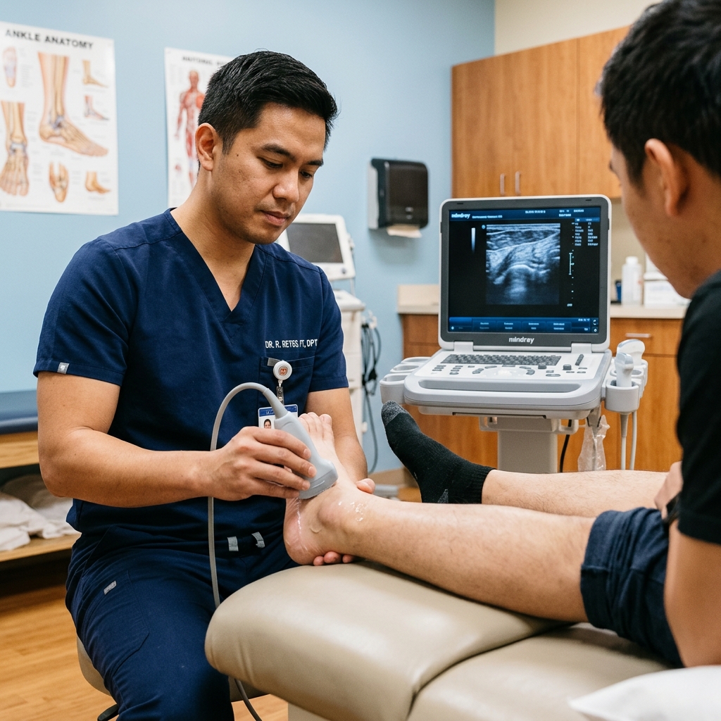
- — Standard X-rays only visualize bones, meaning soft tissue injuries like plantar fasciitis and Achilles tears require high-resolution MSK ultrasound.
- — Bedside MSK ultrasound allows for real-time dynamic stress testing, measuring exact millimeters of tendon thickness and mapping fiber tears in motion.
- — Dosing exercises through progressive mechanical loading is critical for recovery, while aggressive massage or stretching on an undiagnosed tear can worsen the injury.
Understanding First-Step Morning Heel Pain
Morning heel pain represents a common clinical presentation where overnight rest leads to tissue contracture, making the plantar fascia or Achilles tendon highly vulnerable to acute traction forces upon taking the first steps of the day. This first-step pain is primarily driven by plantar fasciitis, insertional Achilles tendinopathy, or acute fascial micro-tears. Because the foot is held in a relaxed, plantarflexed position during sleep, the plantar fascia shortens and begins to heal in a contracted state. When you stand up in the morning, the sudden application of body weight forces the foot into dorsiflexion, abruptly stretching the contracted, inflamed fibers and producing a sharp, tearing sensation.
In my clinical practice, I frequently evaluate patients who present with chronic morning heel pain that has persisted for months. They often describe a severe catching or burning sensation at the bottom of the heel that makes them limp for the first ten to fifteen minutes after waking up. A common source of confusion is having been told that their pain is caused by a "heel spur" seen on a standard X-ray. It is crucial to explain that heel spurs—calcium deposits on the calcaneus—are frequently asymptomatic and are not the primary source of pain. Instead, the real driver is the chronic inflammation, thickening, and micro-tearing of the soft tissues. Bedside musculoskeletal (MSK) ultrasound allows us to look past the bone and directly measure the thickness and structure of the plantar fascia and Achilles tendon, providing immediate diagnostic clarity.
Contrarian Insight 1: The presence of a heel spur on an X-ray is often a clinical distraction. Approximately 10% to 15% of the general population has asymptomatic heel spurs, and many patients with severe plantar fasciitis have no spurs at all. Attributing morning heel pain solely to a spur can lead to inappropriate treatments directed at the bone, whereas the actual pain generator is the mechanical strain and micro-tearing within the soft tissue fascia, which is only visible on high-resolution ultrasound.
To understand the mechanical source of this pain, we must look at the windlass mechanism of the foot. The plantar fascia is a thick band of connective tissue that runs from the heel bone to the base of the toes, acting like a tension cable that supports the medial longitudinal arch. When you take a step, your toes bend upward (dorsiflexion), which winds the plantar fascia around the metatarsal heads, tightening the cable and raising the arch to create a rigid lever for pushing off the ground. If the fascia is thickened or injured, this winding mechanism increases the tensile strain on the calcaneal insertion, triggering sharp pain. High-resolution ultrasound allows us to visualize this insertion point directly, measuring tissue thickness and identifying hypoechoic areas of inflammation or fiber tearing.
Plantar Fasciitis: Visualizing Thickness in Millimeters
Plantar fasciitis is diagnosed on ultrasound when the plantar fascia thickness exceeds 4.0 millimeters at its calcaneal insertion, accompanied by hypoechoic tissue changes and loss of normal fibrillar patterns. Under normal, healthy conditions, the plantar fascia presents as a thin, bright (hyperechoic) band with parallel fibrillar fibers, measuring between 2.0 and 3.0 millimeters in thickness. When chronic strain leads to micro-tearing and failed healing, the body replaces the organized fibers with disorganized, fluid-filled tissue, causing the fascia to swell and thicken.
During a diagnostic ultrasound evaluation, I place the transducer on the plantar aspect of the heel, scanning from the calcaneal tuberosity down the length of the arch. We look for specific sonographic criteria:
- Thickness Cutoff: A plantar fascia thickness greater than 4.0 mm is highly diagnostic of plantar fasciitis. In severe, chronic cases, I frequently measure thickness values exceeding 6.0 or 7.0 mm (Cushman et al., 2025; PMID: 39761995).
- Echogenicity Changes: The normal bright, hyperechoic appearance is replaced by dark, hypoechoic regions, indicating localized swelling and fluid accumulation.
- Fibrillar Disorganization: Loss of the normal parallel fiber alignment, showing a disorganized, amorphous tissue structure.
- Stiffness Mapping (Elastography): Ultrasound elastography reveals a significant decrease in tissue stiffness within the proximal fascia, indicating structural softening and matrix degeneration (Wu et al., 2022; PMID: 35998342).
A systematic review of plantar fascia ultrasound findings confirmed that increased thickness and hypoechoic changes at the calcaneal insertion are the most reliable diagnostic indicators of plantar fasciitis, with thickness showing a direct correlation with symptom severity and walking limitations (Khammas et al., 2023; PMID: 36040577). By measuring these parameters, we can objectively grade the severity of the fasciitis, track the progress of healing during rehabilitation, and identify if a focal tear is present within the fascial fibers, which changes the recovery timeline.
| Pathology | Normal Thickness / State | Ultrasound Diagnostic Cutoff | Dynamic Scan Findings |
|---|---|---|---|
| Plantar Fasciitis | 2.0 to 3.0 mm (thin, bright, parallel fibrillar fibers). | Greater than 4.0 mm at calcaneal insertion with hypoechoic thickening. | Fascial thickening remains static; pain is elicited upon transducer pressure over the medial tubercle. |
| Achilles Tendinopathy | 4.0 to 6.0 mm (uniform tendon thickness, distinct margins). | Greater than 7.0 mm (mild/moderate) or greater than 10.0 mm (severe). | Passive ankle motion reveals localized tendon thickening, neovascularization on power Doppler, and intact sliding. |
| Achilles Tendon Tear | Continuous parallel collagen fibers with zero hypoechoic gaps. | Anechoic gap in fibers, local hematoma, or retracted tendon ends. | Active ankle plantarflexion reveals fiber retraction, gap expansion, or lack of tendon ends approximation. |
| ATFL Ligament Tear (Ankle Sprain) | Uniform thin ligament bridging the fibula and talus bones. | Grade 1: Swollen intact fibers; Grade 2: Disorganized clefts; Grade 3: Complete gap. | Anterior drawer stress test under live scan shows ligament elongation and widening of the talofibular joint gap. |
Achilles Tendon Pain: Differentiating Tendinopathy from Tears
Achilles tendon ultrasound distinguishes tendinopathy (tendon thickness >7.0 mm with disorganized fibers) from acute partial or full-thickness tears (presence of anechoic fiber gaps and fluid collections). The Achilles tendon is the largest and strongest tendon in the body, transmitting the force of the calf muscles to the heel bone to lift the heel during walking and running. Because it is subjected to high mechanical loads, it is prone to chronic degeneration (tendinopathy) and acute structural failure (tears).
During an Achilles ultrasound, I perform both static and dynamic scans. A static scan measures the midportion and insertion of the tendon to check for thickening, which in tendinopathy typically exceeds 7.0 mm (normal is 4.0 to 6.0 mm). In severe tendinopathy, the tendon can expand to over 10.0 mm, showing significant swelling and disorganized, hypoechoic tissue. In contrast, an acute tear presents as a physical disruption of the tendon fibers. Dynamic scanning is critical here: as the patient gently moves their ankle through plantarflexion and dorsiflexion, I can directly visualize the tear gap. If the tear is a partial-thickness tear, some fibers remain intact and slide normally. If it is a full-thickness rupture, I can observe the two retracted ends of the tendon pulling apart, leaving an anechoic (dark), fluid-filled gap where the fibers have separated.
A systematic review and meta-analysis of diagnostic accuracy demonstrated that ultrasound is highly sensitive (exceeding 90%) and specific for diagnosing Achilles tendon ruptures, making it an excellent bedside tool to confirm a rupture immediately during clinical evaluation (Aminlari et al., 2021; PMID: 34801318). Differentiating between tendinopathy and a tear is critical, as a tendinopathy requires a structured, progressive loading rehabilitation program, whereas a full-thickness rupture requires immediate immobilization, orthotic wedge positioning, or surgical referral to prevent permanent calf muscle weakness.
Contrarian Insight 2: I advise my patients that complete pain relief is not an indicator of tendon recovery in chronic Achilles tendinopathy. Chronic tendinosis is characterized by a lack of active inflammation (contrary to the popular term "tendinitis"), meaning that pain can resolve long before the tendon matrix has restored its collagen structure. Progressing to high-impact activities solely because the pain has stopped, without verifying the return of tendon thickness and fiber alignment, often results in recurrent flares or acute rupture.
Sprained Ankle vs. Torn Ligament: Grading Instability
Musculoskeletal ultrasound grades lateral ankle ligament sprains in real-time, distinguishing Grade 1 sprains (swollen, intact fibers) from Grade 2 partial tears and Grade 3 complete ruptures. The lateral ankle ligaments—primarily the anterior talofibular ligament (ATFL) and the calcaneofibular ligament (CFL)—are the most commonly injured structures during an inversion ankle sprain. Standard clinical examinations and X-rays are often insufficient to determine the exact degree of ligament damage, as they cannot visualize soft tissue integrity directly.
To evaluate lateral ankle stability, I perform a dynamic stress ultrasound of the ATFL. While scanning the ligament, I apply a gentle anterior drawer stress to the foot. In a healthy ankle or a Grade 1 sprain, the ligament remains tight and prevents the talus bone from sliding forward. In a Grade 2 partial tear, the ultrasound shows localized hypoechoic clefts within the ligament, and the stress test reveals mild laxity or elongation. In a Grade 3 complete rupture, the ligament is completely torn, and the stress test shows the talus bone sliding abnormally away from the fibula, widening the joint space and demonstrating severe mechanical instability. Research has confirmed that dynamic stress-to-nonstress ATFL length ratios provide clear normative values to diagnose ligament laxity and joint instability objectively (Yokoe et al., 2021; PMID: 34820463). Furthermore, clinical studies have established that MSK ultrasound offers excellent diagnostic value for identifying acute lateral and syndesmotic ankle injuries, matching or exceeding the accuracy of delayed MRI (Baltes et al., 2021; PMID: 33026501).
Contrarian Insight 3: The common practice of obtaining an immediate ankle X-ray after a sprain is useful to rule out fractures, but it can create a false sense of security. A "normal" X-ray report often leads patients and some clinicians to assume that the injury is minor, prompting them to walk on the joint immediately. X-rays cannot visualize ligaments; walking on a Grade 2 or 3 ATFL tear without proper stabilization can cause permanent ligament elongation, leading to chronic ankle instability and recurrent sprains.
Dangers of Aggressive Stretching and Massage on Acute Tears
Aggressive stretching or deep tissue massage on an undiagnosed acute plantar fascia or Achilles tear is clinically contraindicated because the mechanical force can worsen fiber tearing, increase local inflammation, and delay tissue healing. While stretching is a common recommendation for chronic, tight tendons, applying high tensile load or deep pressure to an acute tear disrupts the fragile collagen bridge the body is trying to build, turning a minor partial tear into a complete rupture.
When a patient experiences sudden morning heel pain or Achilles stiffness, their first instinct is often to perform aggressive calf stretches or use a hard roller to massage the bottom of their foot. If the tissue is simply tight from chronic tendinopathy, gentle stretching can be helpful. However, if the patient has a partial plantar fascia tear or acute Achilles tendinitis, the fascia is already micro-torn and highly irritable. Forcing the foot into extreme dorsiflexion pulls the torn ends apart, disrupting the early healing matrix. Similarly, deep massage over the calcaneal insertion applies direct shear force to the healing fibers, causing micro-bleeding and increasing inflammatory pain. In our clinic, we use ultrasound to map the structural state of the tissue first, ensuring we only prescribe stretching when it is safe and appropriate.
From a clinical perspective, we must define the tissue parameters and set clear safety boundaries:
- Tissue State: The target tissue state is a thickened plantar fascia or Achilles tendon (indicative of tendinopathy) with intact, continuous fibers and minimal acute inflammatory fluid. If ultrasound reveals a focal hypoechoic cleft (a tear) or an active hematoma, high-tensile stretching must be avoided.
- Contraindication Logic: Aggressive stretching, eccentric loading, and deep manual therapy are strictly contraindicated in patients with acute partial-thickness or full-thickness tears, suspected infection, or severe acute inflammation (gouty flare or active synovitis). Stretching a torn tendon pulls the healing margins apart, preventing proper cross-linking of collagen fibers.
- Progression Criteria: The patient progresses from protection and gentle active range of motion to progressive loading once the ultrasound shows tissue continuity (resolution of hypoechoic clefts) and the patient can perform pain-free calf raises and walk without a limp.
- Physiological Rationale: Healing tendons and fascia require controlled mechanical loading (tension) to stimulate tenocytes to produce new type I collagen. However, the load must be dosed to the tissue's capacity. During the early healing phase, relative rest and orthotic offloading allow the initial collagen scaffold to form. Once stable, progressive loading guides the alignment of these new fibers along the lines of mechanical stress, restoring tensile strength.
Evidence-to-Practice: Loading Protocols for Heel and Ankle Injuries
In translating clinical evidence to real-world recovery, we must bridge the gap between ideal research protocols and the practical realities patients face. Modern clinical literature proves that foot and ankle injuries respond best to structured, progressive mechanical loading rather than prolonged rest or aggressive stretching. Ankle ligament studies demonstrate that early functional mobilization using a supportive brace achieves superior long-term stability and faster return to activity compared to prolonged cast immobilization (Baltes et al., 2021; PMID: 33026501). Similarly, systematic reviews of plantar fasciitis confirm that progressive loading protocols (using a towel roll under the toes to engage the windlass mechanism during calf raises) significantly improve fascia tissue density and reduce morning pain (Khammas et al., 2023; PMID: 36040577). While ideal protocols advocate for supervised, twice-weekly physical therapy sessions for 12 weeks to guide this loading, this schedule is often unrealistic for patients due to work demands, travel constraints, and cost sensitivity.
To address these real-world barriers, I implement a practical, staged care pathway in our Vigan clinic. During the initial consultation, we perform a bedside MSK ultrasound to confirm the diagnosis and measure the tissue thickness. If burning or tingling dominates rather than morning mechanical heel pain, we also screen for nerve compression using our clinical approach for Tarsal Tunnel Syndrome. If the plantar fascia is thickened but intact (>4.0 mm), we review the dynamic windlass mechanism on the screen and teach the patient a targeted home exercise program during the same visit. The program focuses on pain-free, low-load progressive loading (such as isometric calf raises with toe support) and guided heel support. To address the financial and travel barriers of frequent clinic visits, the patient performs these exercises at home 2 times daily for 6 weeks, utilizing accessible home items and wearing supportive indoor footwear instead of walking barefoot on hard floors. We monitor their safety and progress through weekly digital or telephone check-ins, tracking safety signs such as worsening morning pain, new calf swelling, or loss of ankle motion. The patient only returns to the clinic for a follow-up ultrasound assessment at the 6-week mark, or is escalated for advanced care (such as custom orthotic fitting or PRP therapy) if they fail to show objective functional improvement after consistent home program adherence, ensuring a safe, realistic, and highly effective pathway to recovery.
Warning Signs: When Foot and Ankle Pain Requires Urgent Care
Immediate clinical evaluation is indicated when foot or ankle pain is accompanied by signs of acute structural failure, infection, or vascular compromise. While most plantar fascia and Achilles tendon issues resolve with structured non-surgical care, certain clinical presentations require urgent specialist assessment to prevent permanent disability. Delaying evaluation in these cases can lead to chronic instability, tendon retraction, or severe systemic complications.
Please seek immediate medical assessment if you experience any of the following warning signs:
- The "Pop" and Drop: A sudden, audible "pop" in the back of the heel accompanied by a sharp pain and an immediate inability to push off your foot or raise your heel. This is a classic sign of an acute Achilles tendon rupture.
- Visible Tendon Deformity: A visible gap or hollowing in the back of your ankle above the heel bone, which suggests a retracted tendon rupture.
- Severe Calf Swelling: Progressive pain, swelling, and redness in your calf muscle after an ankle sprain, which can indicate a deep vein thrombosis (blood clot) rather than a simple sprain.
- Fever and Localized Heat: Severe, throbbing foot pain accompanied by a fever, chills, or spreading redness and warmth, pointing to a bone or joint infection.
Contrarian Insight 4: I warn my patients that the complete resolution of pain does not mean the tissue has fully recovered. In chronic plantar fasciitis and Achilles tendinopathy, the pain can subside after rest or anti-inflammatory treatments while the tissue remains structurally weak and thickened. Returning to running or heavy labor without a guided, progressive strengthening program to restore the tissue's load tolerance often results in recurrent, worse injuries.
In our clinic, our primary goal is to help you understand your symptoms and make informed decisions about your recovery. If your morning heel pain or ankle stiffness does not improve after 3 to 4 weeks of rest and supportive shoe wear, a proper diagnostic assessment is necessary. Using bedside MSK ultrasound, we can immediately visualize your tendons and ligaments, identify whether the issue is tendinopathy, a tear, or chronic ligament laxity, and build a safe, non-surgical treatment plan that fits your life and helps you return to activity comfortably and safely.
References & Clinical Evidence
- [1] Baltes TPA, Arnáiz J, Geertsema L, Geertsema C, D'Hooghe P, Kerkhoffs GMMJ, Tol JL. Diagnostic value of ultrasonography in acute lateral and syndesmotic ligamentous ankle injuries. Eur Radiol. 2021 May;31(5):3130-3138. doi: 10.1007/s00330-020-07305-7. PMID: 33026501.
- [2] Aminlari A, Stone J, McKee R, Subramony R, Nadolski A, Tolia V, Hayden SR. Diagnosing Achilles Tendon Rupture with Ultrasound in Patients Treated Surgically: A Systematic Review and Meta-Analysis. J Emerg Med. 2021 Nov;61(5):540-547. doi: 10.1016/j.jemermed.2021.09.008. PMID: 34801318.
- [3] Cushman DM, Stokes D, Vu L, Corcoran B, Fredericson M, Eby SF, Teramoto M. Ultrasound as a predictor of time-loss injury for the patellar tendon, Achilles tendon and plantar fascia in division I collegiate athletes. Br J Sports Med. 2025 Jan 14;59(1):15-21. doi: 10.1136/bjsports-2024-109066. PMID: 39761995.
- [4] Wu CH, Chiu YH, Chang KV, Wu WT, Özçakar L. Ultrasound elastography for the evaluation of plantar fasciitis: A systematic review and meta-analysis. Eur J Radiol. 2022 Oct;155:110495. doi: 10.1016/j.ejrad.2022.110495. PMID: 35998342.
- [5] Khammas ASA, Mahmud R, Abu Hassan H, Ibrahim I, Mohammed SS. An assessment of plantar fascia with ultrasound findings in patients with plantar fasciitis: a systematic review. J Ultrasound. 2023 Mar;26(1):1-10. doi: 10.1007/s40477-022-00712-0. PMID: 36040577.
- [6] Yokoe T, Tajima T, Kawagoe S, Yamaguchi N, Morita Y, Chosa E. The Ratio of Stress to Nonstress Anterior Talofibular Ligament Length on Ultrasonography: Normative Values. Orthop J Sports Med. 2021 Nov 16;9(11):23259671211056305. doi: 10.1177/23259671211056305. PMID: 34820463.
* Clinical references are provided to support the medical claims made in this article. TeraCare adheres to evidence-based practices in physical medicine and rehabilitation.

Dr. Ben Rabara
Dr. Ben Rabara is a Board-Certified Physiatrist specializing in Physical Medicine and Rehabilitation. He focuses on non-surgical, precision treatments for musculoskeletal conditions, utilizing advanced diagnostics like MSK Ultrasound.
Medical Disclaimer: The information provided in this article is for educational purposes only and does not substitute for professional medical advice, diagnosis, or treatment. Always consult a qualified physician for your specific health conditions.