- — Musculoskeletal (MSK) ultrasound in the Philippines costs a fraction of an MRI, typically ₱3,000 for a formal radiological report and free for bedside assessment during clinical decision-making.
- — bedside ultrasound is dynamic, allowing the physician to view muscles, tendons, and ligaments in active motion to detect impingements that static MRIs miss.
- — Bedside scanning eliminates waiting lists and patient claustrophobia, offering immediate diagnostic results during your initial consultation.
- — Delaying a diagnosis with traditional massage (hilot) or tight bandages (benda) can lead to permanent muscle atrophy or tendon rupture.
When you experience persistent joint or tendon pain in the Philippines, the clinical path to recovery depends entirely on obtaining an accurate, timely diagnosis. Too often, patients are left choosing between two extremes: waiting weeks for a highly expensive, claustrophobia-inducing Magnetic Resonance Imaging (MRI) scan at a major regional hospital, or attempting traditional, unguided remedies that can worsen a hidden tear. In my practice at Rabara Rehabilitation in Vigan, Ilocos Sur, I use musculoskeletal (MSK) ultrasound as a point-of-care diagnostic tool during consultations. This bedside imaging modality provides immediate clinical answers, allows for dynamic joint evaluation, and eliminates the substantial wait times and cost barriers associated with traditional static imaging.
Diagnostic MSK Ultrasound vs. MRI: Cost and Key Differences
Joint diagnostic imaging costs vary widely in the Philippines, with a standard MRI ranging from ₱10,000 to ₱20,000+ depending on the facility and the need for contrast agents, while a point-of-care musculoskeletal (MSK) ultrasound at our Vigan clinic is ₱3,000 for an official report (and is included for free during physical medicine consultations if no formal written report is required). This price gap makes ultrasound the most accessible first-line choice for soft-tissue diagnostics, allowing patients to bypass the financial strain of larger hospital visits. While an MRI remains the gold standard for visualizing deep intra-articular structures like the anterior cruciate ligament (ACL) or meniscus within the knee cavity, high-resolution ultrasound provides equal or superior clarity for superficial structures, including the rotator cuff tendons, superficial ligaments, and fluid collection.
The difference in cost is rooted in the underlying technology and facility overhead. MRI machines are multi-million peso installations requiring specialized shielding, liquid helium cooling, and substantial electrical power. These costs are passed directly to the patient. Conversely, MSK ultrasound utilizes high-frequency sound waves (ranging from 5 to 18 MHz depending on the target tissue depth) generated by a compact transducer. This transducer transmits sound waves into the tissue, which reflect off anatomical boundaries to create real-time, millimeter-resolution images of muscles, tendons, ligaments, nerves, and joints. Extensive clinical literature demonstrates that for superficial soft tissues, ultrasound exhibits excellent diagnostic agreement with MRI. For instance, in a tertiary care evaluation of rotator cuff pathology, high-resolution ultrasound demonstrated high diagnostic accuracy and sensitivity matching MRI for full-thickness tears, confirming its role as a premier cost-effective screening modality (Pandya et al., 2025; PMID: 41846768). Additionally, comparative diagnostic assessments show that ultrasound provides a sensitivity of up to 95% and specificity exceeding 90% for detecting subacromial-subdeltoid bursitis and joint effusion (Madhavi et al., 2024; PMID: 39350841). This means that for conditions like shoulder impingement or swollen knees, spending ₱15,000 on an MRI yields no additional diagnostic information over a targeted ₱3,000 ultrasound scan.
| Diagnostic Feature | Point-of-Care MSK Ultrasound | Standard Joint MRI |
|---|---|---|
| Average Cost | ₱3,000 (Or free in clinical consultation) | ₱10,000 – ₱20,000+ |
| Waiting Time | Immediate (Performed during consultation) | 4 to 8 weeks (In public/regional centers) |
| Claustrophobia Risk | Zero (Comfortable open-room scan) | High (Up to 15% abort or panic rate) |
| Assessment Type | Dynamic (Joint evaluated in active motion) | Static (Lying completely still inside tube) |
| Radiation Exposure | Zero (Safe high-frequency sound waves) | Zero (Magnetic fields) |
| Primary Diagnostics | Tendon tears, bursa swelling, muscle sprains, effusions | Deep ACL, meniscus, bone marrow edema, internal joint cavities |
Why Dynamic Joint Imaging Outperforms Static Scans
Dynamic musculoskeletal ultrasound allows the physician to move the joint in real-time under active scanning, whereas MRI requires the patient to remain perfectly still in a static position. This dynamic movement reveals snapping tendons, sliding ligament fibers, and mechanical impingements that static images completely miss. By observing the patient's joints during active contraction and passive stress testing, I can directly correlate their physical pain symptoms with the visual displacement of anatomical structures. This real-time feedback is an invaluable clinical resource that static cross-sectional imaging cannot replicate.
In a standard static scan, the joint is imaged in a neutral, non-weight-bearing position. This static approach can hide pathologies that only manifest during specific arcs of motion. For example, in patients presenting with shoulder pain (masakit na balikat), a rotator cuff tendon may appear structurally intact when resting at the patient's side. However, when I perform a dynamic ultrasound scan, I can guide the patient's arm into abduction and internal rotation (mimicking the painful movement). Under the ultrasound transducer, we can watch the supraspinatus tendon slide beneath the acromion process. If there is dynamic impingement, the tendon or the subacromial-subdeltoid bursa will visibly bunch up or catch against the bone, directly reproducing the patient's pain on a scale of 0 to 10. This dynamic sliding window evaluation allows me to instantly identify subacromial friction, transient subluxations, and unstable tendon micro-tears that flatten out when the limb is at rest (Pandya et al., 2025; PMID: 41846768). Furthermore, for knee diagnostics, dynamic scanning allows us to stress-test the collateral ligaments (such as the medial collateral ligament) or observe the patellar tendon gliding through its groove, identifying mechanical catching, ligament laxity, or joint subluxations that static imaging fails to capture.
Beyond the Cost: Bypassing MRI Claustrophobia and Waiting Lists in Vigan
Musculoskeletal ultrasound resolves the two biggest clinical bottlenecks in provincial healthcare: patient claustrophobia from enclosed MRI tubes and weeks-long hospital waiting lists. Because ultrasound is performed bedside at point-of-care, patients receive immediate diagnostic clarity without anxiety or delay. This rapid diagnostic turnaround is critical for preventing acute sports injuries or work-related joint strains from turning into chronic, poorly managed disabilities due to delayed clinical decisions.
For many patients in Vigan and the wider Ilocos Sur province, obtaining an MRI is not just a financial burden, but an operational logistical hurdle. The closest open MRI machines may require a 1.5-hour to 2-hour drive to Ilocos Norte or La Union, which is difficult for elderly patients or those with severe mobility limitations. Furthermore, enclosed MRI scanners force the patient to lie inside a narrow, loud tube for 30 to 45 minutes. Up to 15% of patients experience significant claustrophobia or panic attacks during the procedure, occasionally requiring sedation or leading to aborted scans. Bedside MSK ultrasound bypasses this entirely. The scan is performed in an open, comfortable consultation room with a family member present. In addition, the wait times for diagnostic imaging in government tertiary hospitals can stretch from 4 to 8 weeks due to high patient volume. In contrast, on-site MSK ultrasound within the clinic provides diagnostic clarity during the initial visit itself. Studies evaluating on-site ultrasound in outpatient clinics have shown that this point-of-care integration avoids unnecessary external imaging referrals, reduces diagnostic delay, and saves significant health-system costs (Acebes et al., 2021; PMID: 33140101). By using ultrasound as a triage tool, we can rapidly scan a painful joint and determine if the patient has a simple sprain that can be managed conservatively, or if they have a complex intra-articular tear that justifies the cost and wait time of an MRI (Awan et al., 2024; PMID: 39451466).
The Danger of the "Wait-and-See" or "Hilot-First" Trap
Treating a joint sprain or tendon tear with standard compression bandages or traditional massage without prior imaging can turn a simple micro-tear into permanent instability. Accurate ultrasound diagnostics prevent this delay, directing patients to the correct physiological loading protocol rather than prolonged, harmful rest. When a patient in the Ilocos region suffers a joint injury (pilay), the common cultural response is to apply a tight elastic bandage (benda sa pilay) and wait for weeks, or visit a traditional practitioner (manghihilot) for vigorous massage. Without a structural diagnosis, these approaches can compromise joint function.
This "wait-and-see" approach is based on the outdated assumption that all soft-tissue injuries simply require rest to heal. However, tendons and ligaments have a poor blood supply and do not heal efficiently through complete immobilization. For instance, if a patient has a partial tear of the patellar tendon or the Achilles tendon, resting the limb for several weeks causes the surrounding muscle fibers to atrophy and decreases the tendon's mechanical load-bearing capacity. When the patient eventually returns to activity, the weakened tendon cannot handle the sudden tension, leading to recurrent micro-tearing and chronic tendinosis. Conversely, aggressive traditional manipulation (hilot) applied to an undiagnosed tendon tear or an inflamed bursa can physically worsen the structural damage, occasionally causing a partial tear to progress to a full-thickness rupture. Soft-tissue ultrasound acts as a rapid, high-accuracy screening method to identify tendon ruptures and guide early active rehabilitation (Crouch-Smith et al., 2026; PMID: 39502676). By using ultrasound to visualize the exact tissue state, I can design a progressive loading rehabilitation program (typically spanning 6 to 12 weeks) that applies safe, controlled mechanical tension to stimulate collagen alignment and restore tendon tensile strength. This ensures that the patient does not waste months in a state of chronic pain and functional decline.
What to Expect During an MSK Ultrasound in Our Vigan Clinic
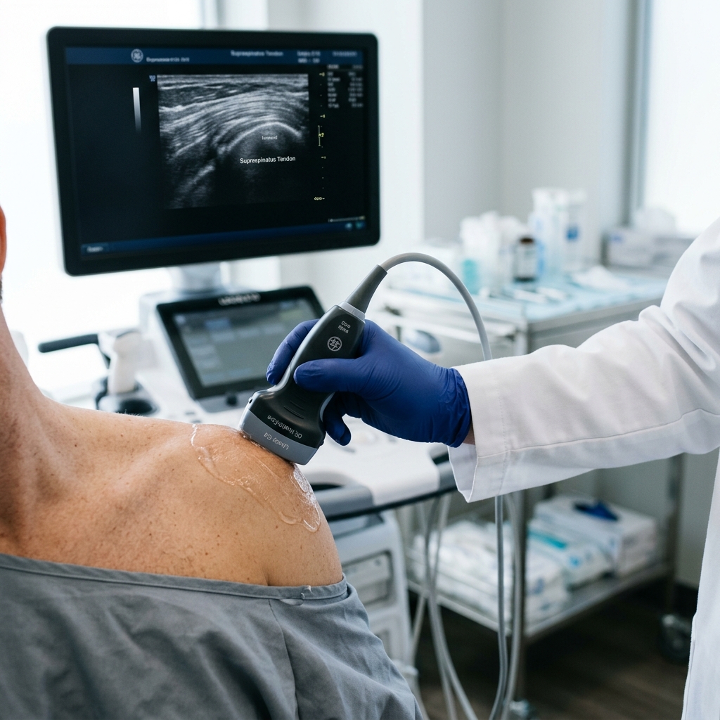
Performing a bedside musculoskeletal ultrasound takes 15 to 30 minutes, during which a conductive gel is applied and a hand-held probe scans the joint in real-time. The patient feels no discomfort, views the live sonogram, and receives an immediate diagnostic interpretation from the physician. Unlike ionizing radiation methods like X-rays, ultrasound utilizes safe acoustic energy, meaning there is zero radiation exposure and the scan can be repeated safely to monitor tissue healing over time.
When you arrive at our clinic in Vigan for a joint evaluation, the process is streamlined and interactive. The diagnostic workflow follows these steps:
- Clinical History and Physical Examination: I first evaluate the joint's range of motion, perform specialized orthopedic stress tests, and map the patient's pain points.
- Gel Application: A water-soluble, hypoallergenic conductive gel is applied to the skin over the target joint (such as the shoulder, knee, or ankle). This gel eliminates air pockets between the transducer and the skin, allowing the sound waves to travel clearly into the body.
- Real-Time Scanning: I place the high-resolution linear transducer over the target structures. I will scan the tissues in two planes (longitudinal and transverse) to evaluate the fibers in their entirety. During the scan, I will ask you to gently contract your muscles or move your joint so we can observe the sliding mechanics of the tendons.
- Immediate Discussion: You can look at the ultrasound monitor while the scan is occurring. I will point out the key structures, such as the supraspinatus tendon, the subacromial bursa, or the joint space, explaining what is normal and where inflammation or tearing is present.
- Clinical Decision-Making: Once the scan is complete, we discuss the findings immediately. If there is a significant fluid collection (effusion) causing severe pressure and pain, we can perform an immediate ultrasound-guided aspiration to drain the fluid, followed by an target rehabilitation program.
PhilHealth, HMOs, and Private Health Coverage for Joint Ultrasound
Outpatient MSK ultrasound and physical medicine consultations qualify for senior citizen and PWD discounts under Philippine law, reducing overall diagnostic costs. While PhilHealth coverage varies for outpatient diagnostic imaging, private HMOs often reimburse ultrasound scans when ordered by a specialist. Understanding these health-system mechanics helps patients manage their outpatient expenses without sacrificing the quality of their diagnostic care.
For patients utilizing private health insurance (HMOs) or seeking reimbursement, the diagnostic request must be officially ordered by a specialist (such as a Physiatrist or Orthopedist) to qualify for coverage. When you receive a consultation at our clinic, I provide the necessary medical certificates, diagnostic codes, and official ultrasound reports required by insurance companies. For senior citizens and Persons with Disabilities (PWDs), a 20% discount and 12% Value Added Tax (VAT) exemption are applied to both the consultation fee and the official diagnostic ultrasound report fee, as mandated by the Philippine government. It is important to note that while government hospitals may offer slightly lower nominal fees for ultrasound scans, the hidden costs of travel, long waiting times, and delayed treatment decisions often outweigh the minor price difference. By choosing a point-of-care outpatient scan, you get immediate answers, allowing you to start the correct treatment plan on the same day and avoid the costly complications of chronic joint degeneration.
If you prefer a Taglish pricing guide for specific body parts, see our magkano ultrasound sa joints, balikat, at kamay page.
References & Clinical Evidence
- [1] Pandya S, et al. Comparative Analysis of Ultrasound and MRI in Diagnosing Rotator Cuff Injuries in a Tertiary Care Setting. J Pharm Bioallied Sci. 2025. PMID: 41846768. DOI: 10.4103/jpbs.jpbs_1527_25.
- [2] Madhavi P, et al. Diagnostic Accuracy of USG and MRI for the Detection of Rotator Cuff Injury. Cureus. 2024. PMID: 39350841. DOI: 10.7759/cureus.68199.
- [3] Awan F, et al. The Utility of a Community-Based Knee Ultrasound in Detecting Meniscal Tears: A Retrospective Analysis in Comparison with MRI. Healthcare (Basel). 2024. PMID: 39451466. DOI: 10.3390/healthcare12202051.
- [4] Acebes C, et al. Cost-effectiveness of on-site musculoskeletal ultrasound in an outpatient rheumatology clinic. Rheumatology (Oxford). 2021. PMID: 33140101. DOI: 10.1093/rheumatology/keaa678.
- [5] Crouch-Smith HA, et al. A retrospective study comparing ultrasound and MRI for diagnosing quadriceps and patellar tendon ruptures, along with a cost-benefit analysis of these imaging techniques. Journal of Orthopaedics. 2026. PMID: 39502676. DOI: 10.1016/j.jor.2025.11.015.
* Clinical references are provided to support the medical claims made in this article. TeraCare adheres to evidence-based practices in physical medicine and rehabilitation.

Dr. Ben Rabara
Dr. Ben Rabara is a Board-Certified Physiatrist specializing in Physical Medicine and Rehabilitation. He focuses on non-surgical, precision treatments for musculoskeletal conditions, utilizing advanced diagnostics like MSK Ultrasound.
Medical Disclaimer: The information provided in this article is for educational purposes only and does not substitute for professional medical advice, diagnosis, or treatment. Always consult a qualified physician for your specific health conditions.