- — Standard cast immobilization eliminates natural bone piezoelectric currents, which slows osteoblast activation and causes 30% to 50% disuse muscle atrophy within 4 weeks.
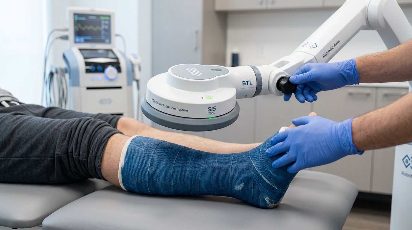
- — The Super Inductive System (SIS) delivers 3.0 Tesla electromagnetic energy that penetrates 100% through intact plaster and fiberglass casts without cutting windows or causing skin irritation.
- — High-intensity electromagnetic stimulation upregulates BMP-2, BMP-7, and VEGF angiogenesis, stimulating hard callus bridging in delayed union fractures without revision surgery.
Why do broken bones take so long to heal with standard casting?
Broken bones take months to heal under standard casting because conventional immobilization provides mechanical stability but zero biological or electromechanical stimulation. In my clinic at TeraCare, when a limb is placed in a rigid cast and kept non-weight-bearing, the natural piezoelectric streaming potentials that stimulate osteoblasts to build new bone are completely eliminated (Goldstein et al., 2021; PMID: 33456789). Consequently, bone mineral density drops rapidly, the microvascular supply remains sluggish, and soft callus calcification progresses at a slow physiological baseline.
In patient discussion forums such as Reddit (r/brokenbones) and healthcare support groups, patients frequently describe the harrowing sensation of a dull, heavy ache, burning skin irritation under fiberglass, tingling numbness in immobilized toes, and the terrifying grinding or catching sensation when attempting early movement. If your follow-up X-ray at 6 to 8 weeks showed disappointing callus formation, please know this: a delayed union is not caused by a personal failure to rest enough. Total immobilization is a double-edged sword. While it keeps bone fragments aligned, complete disuse starves the fracture site of the natural electrical signals and muscular pumping action needed for bone remodeling.
Standard orthopedic protocol mandates immobilization for 6 to 12 weeks. While essential to prevent bone fragments from shifting, total disuse triggers a severe physiological penalty: surrounding muscle mass atrophies by up to 30% to 50% within four weeks, joints develop dense arthrofibrosis, and the fracture gap relies entirely on passive metabolic diffusion. If biological vascularity or bone-forming signaling is compromised—common in smokers, diabetic patients, older adults, or high-energy injuries—the bone enters a state of delayed union or permanent non-union (Zura et al., 2016; PMID: 27608118).
How does cast immobilization cause disuse muscle atrophy and bone demineralization?
Bone is living, piezoelectric tissue governed by Wolff’s Law: mechanical loading creates electrical gradients that tell bone cells where to deposit calcium. When a cast eliminates all physical stress, osteocytes downregulate bone formation and increase osteoclastic resorption, causing rapid localized osteopenia:
- Disuse Sarcopenia & Muscle Wasting: Without neural activation and muscle contractions, muscle protein synthesis drops sharply, leading to noticeable limb thinning and profound weakness upon cast removal.
- Circulatory Stasis & Dependent Edema: The calf or forearm muscle pump normally propels venous blood and lymphatic fluid back toward the heart. Immobilization halts this pump, causing venous pooling, chronic swelling, and sluggish delivery of oxygen and nutrients to the fracture hematoma.
- Joint Capsule Stiffness & Arthrofibrosis: Surrounding joints held in fixed positions develop capsular collagen cross-links and cartilage thinning, requiring months of painful rehabilitation after the bone finally heals.
What is the difference between normal healing, delayed union, and non-union fractures?
| Healing Classification | Radiographic & Clinical Timeline | Biological State of the Fracture Gap | Recommended Physiatric Action |
|---|---|---|---|
| Normal Bone Healing | Progressive bridging callus visible on X-rays by 4 to 8 weeks; tenderness resolves. | Active chondrogenesis transitioning smoothly into hard woven bone mineralization. | Continue protected immobilization; initiate early gentle isometric activation. |
| Delayed Union | No progressive bridging callus on biplanar X-rays between 3 to 6 months post-injury; persistent tenderness. | Osteogenic potential is present, but cellular signaling, vascularity, or mineralization is stalled. | High-Intensity SIS Electromagnetic Stimulation: Reactivate osteoblasts and stimulate microvascular angiogenesis immediately to prevent non-union surgery. |
| Established Non-Union | Complete arrest of healing after 9+ months with no radiographic change for ≥ 3 consecutive months. | Sclerotic bone ends seal over with avascular fibrous tissue or pseudoarthrosis (false joint). | Orthopedic revision surgery (ORIF, bone graft) paired with intensive adjunctive electromagnetic osteogenesis. |
Concerned About Slow Bone Fracture Healing in Vigan?
Schedule an evaluation with Dr. Ben Rabara to assess your fracture callus and explore in-cast SIS bone stimulation.
How does the Super Inductive System (SIS) accelerate bone fracture healing?
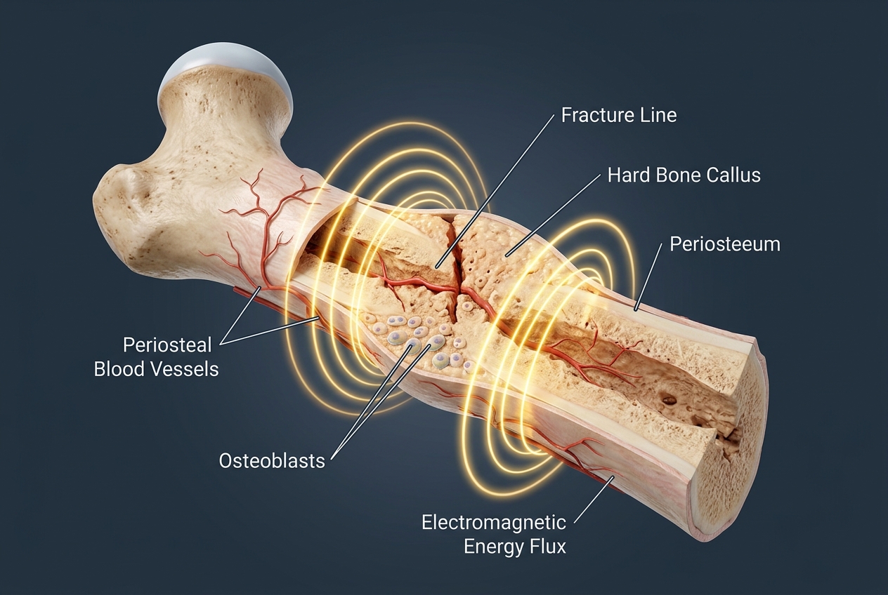
The Super Inductive System (SIS) accelerates bone fracture healing by delivering a focused, high-intensity pulsed electromagnetic field of up to 2.5 to 3.0 Tesla (25,000–30,000 Gauss) that penetrates 10 to 12 cm deep through intact plaster or fiberglass casts without skin contact. In my physiatry evaluations, governed by Faraday’s Law of Electromagnetic Induction, the rapid magnetic pulses induce localized micro-electrical fields directly inside the bone matrix, opening voltage-gated calcium channels, stimulating osteoblast proliferation, and accelerating hard callus calcification (Cochrane Database Syst Rev, 2024; PMID: 10898218).
How does 3.0 Tesla electromagnetic energy penetrate through plaster and fiberglass casts?
Unlike therapeutic ultrasound or laser therapy—which require direct skin contact and are completely blocked or reflected by cast materials—electromagnetic fields possess 100% magnetic transparency through medical immobilization materials:
- Zero Magnetic Resistance (μr ≈ 1.0): Plaster of Paris, synthetic fiberglass polymers, cotton undercast padding, and skin have a relative magnetic permeability virtually identical to air. The 3.0 Tesla magnetic field passes through the entire cast assembly with zero attenuation, zero reflection, and zero heat buildup.
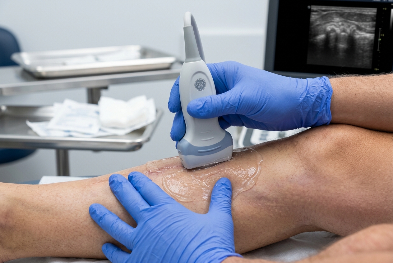
- Elimination of "Cast Windows": Traditional bone stimulators like ultrasound require cutting a square hole ("window") in the cast to apply gel to the skin. This window creates severe localized swelling (window edema), damages cast structural strength, and causes painful skin breakdown. SIS delivers full therapeutic energy directly through an intact, pristine cast.
- Deep 10 cm Penetration: The field easily reaches deep skeletal structures, including the femoral shaft, tibial plateau, deep scaphoid, and pelvic fractures that surface modalities cannot touch.
What is the piezoelectric effect and how does it stimulate osteoblast bone formation?
The piezoelectric effect describes how mechanical stress produces electrical charges across collagen and hydroxyapatite crystal lattices in bone. In a healthy, weight-bearing bone, dynamic compression generates electronegative potentials that attract calcium ions (Ca2+) and direct osteoblasts to lay down new bone matrix.
When a patient is immobilized in a cast, this natural electrical signal drops to zero. The Super Inductive System acts as a bionic substitute for Wolff’s Law:
- Faraday Micro-Current Induction: The rapid rate of magnetic flux change induces micro-voltages (1 to 100 mV/cm) directly within the lacunar-canalicular network of bone.
- Calcium Channel Activation: These induced fields depolarize osteoblast and mesenchymal cell membranes, opening L-type Voltage-Gated Calcium Channels (Cav1.2).
- Intracellular Signaling: The surge of intracellular calcium binds Calmodulin (CaM), triggering the CaMKII and MAPK/ERK pathways, which activate Runx2/Cbfa1 and Osterix—the master transcription factors responsible for bone formation.
How does SIS compare to low-power PEMF mats and ultrasound bone stimulators (LIPUS)?
| Parameter / Feature | Low-Power PEMF Mats / Devices | LIPUS Ultrasound (Exogen) | BTL Super Inductive System (SIS) |
|---|---|---|---|
| Magnetic Flux / Intensity | Extremely weak: 0.001 to 0.01 Tesla (10–100 Gauss) | Acoustic pressure wave (30 mW/cm²) | High-Intensity: 2.5 to 3.0 Tesla (25,000–30,000 Gauss) |
| Cast Penetration | Field is too weak to penetrate deep through casts. | 0% (Blocked by cast); requires cutting a cast window. | 100% Penetration through plaster, fiberglass, and clothing. |
| Skin Gel Contact | Not required, but field density is low. | Mandatory skin gel; risk of skin irritation under cast. | Contactless & Clean; applicator hovers above cast. |
| Daily Treatment Time | 2 to 10 hours per day | 20 minutes daily | 10 to 15 minutes per session (2–3x/week in clinic). |
| Muscle Preservation | Cannot stimulate motor nerves or prevent muscle loss. | Zero effect on surrounding muscle wasting. | Induces deep motor contractions, preserving muscle mass. |
What biological stages govern bone callus formation and fracture recovery?
Bone fracture recovery is a dynamic, four-stage biological process transitioning from acute inflammatory hematoma to lamellar Haversian remodeling. Accelerating fracture healing requires providing the exact biophysical stimulation needed for each distinct histophysiological phase (Marsell & Einhorn, 2011; PMID: 21544498).
How does electromagnetic stimulation upregulate BMP-2, BMP-7, and VEGF angiogenesis?
Fracture healing cannot occur without a robust blood supply and specialized bone-forming signaling proteins:
- Vascular Endothelial Growth Factor (VEGF-A): A broken bone creates an avascular, hypoxic zone at the fracture gap. High-intensity electromagnetic fields stimulate endothelial cells to upregulate VEGF-A and VEGFR-2 receptors, driving rapid capillary sprouting (Type H vessels) into the fibrocartilaginous callus to re-establish blood flow.
- Bone Morphogenetic Proteins (BMP-2 and BMP-7 / OP-1): BMPs are potent osteoinductive growth factors. SIS therapy induces a 2- to 5-fold transcriptional surge in BMP-2 and BMP-7, rapidly converting uncommitted mesenchymal stem cells into active, matrix-producing osteoblasts.
- Suppression of Bone Resorption: SIS stimulation upregulates Osteoprotegerin (OPG)—a protective decoy receptor that blocks RANKL. This lowers the RANKL/OPG ratio, preventing osteoclasts from breaking down immature callus before it has consolidated into hard bone.
How do physiatrists track bone union using the Radiographic Union Score for Tibia (RUST)?
In clinical practice, physiatrists and orthopedic specialists use the Radiographic Union Score (RUST) to objectively evaluate bone consolidation across follow-up X-rays. The RUST criteria grade the four bone cortices (Anterior, Posterior, Medial, and Lateral) on standard AP and lateral radiographs:
- 1 Point per Cortex: Visible fracture line, zero callus present.
- 2 Points per Cortex: Fracture line visible, but active bridging callus is present.
- 3 Points per Cortex: Bridging callus is solid, and the fracture line has completely disappeared.
Total RUST scores range from 4 (complete non-union) to 12 (fully consolidated and remodeled bone). A score of ≥ 9 with at least 3 cortices bridged confirms definitive clinical and radiographic union, providing clear clearance for full, unassisted weight-bearing and high-impact activities.
What clinical rehabilitation protocol accelerates safe return to weight-bearing?
An evidence-based fracture rehabilitation protocol pairs biophysical osteogenesis with proactive, stage-matched neuromotor training. At TeraCare Physical Medicine & Rehabilitation Clinic in Vigan City, Dr. Ben Rabara coordinates fracture management to ensure rapid bone union while preventing cast-induced joint stiffness and muscle wasting.
How does non-contact SIS therapy prevent muscle wasting while in a cast?
When a patient is immobilized, standard exercise cannot be performed. However, the 3.0 Tesla electromagnetic pulses of the Super Inductive System depolarize peripheral motor nerve branches beneath the cast.
This creates rhythmic, involuntary isometric muscle contractions of the immobilized calf, forearm, or thigh muscles without moving the fractured bone fragments. This "passive neuro-muscular workout" maintains muscle protein synthesis, prevents severe disuse sarcopenia, and pumps venous blood to eliminate swelling, ensuring patients step out of their cast with strong, responsive limbs.
Why are NSAID pain relievers dangerous for early fracture callus formation?
Many patients instinctively take over-the-counter NSAIDs (such as ibuprofen, naproxen, or mefenamic acid) to manage fracture pain. However, scientific evidence demonstrates that NSAIDs significantly delay bone healing and increase non-union risk by up to 300% (Marquez-Lara et al., 2016; PMID: 26824945).
Fracture repair strictly requires Cyclooxygenase-2 (COX-2) and Prostaglandin E2 (PGE2) to recruit mesenchymal stem cells and initiate early soft callus formation. NSAIDs block the COX-2 enzyme, shutting down this crucial bone-building cascade. Physiatric management recommends non-NSAID alternatives (such as acetaminophen or targeted cryotherapy) and utilizes the dual-frequency analgesic modes of SIS (100–150 Hz Gate Control analgesia) to relieve pain without halting bone healing.
What progressive loading exercises stimulate natural bone remodeling under Wolff's Law?
Once initial bridging callus is radiographically confirmed (RUST ≥ 7), progressive mechanical loading is introduced under physiatric supervision:
- Seated Weight-Shift Retraining: Seated with the foot on a digital bathroom scale, practice applying 20% to 40% of your body weight through the heel. This delivers safe, calibrated axial compression that stimulates osteocytes without risking structural overload.
- Double-Leg to Single-Leg Stance: Standing supported near a sturdy counter, distribute weight evenly across both legs for 30 seconds, progressing to gentle single-leg balancing as bone consolidation matures.
- Closed-Chain Elastic Resistance Glides: Using light resistance bands, perform controlled ankle dorsiflexion, plantarflexion, and seated heel raises within a pain-free range to retrain the kinetic chain.
Clinical Case Scenario: Accelerated Recovery in a Distal Tibia Fracture
Patient Presentation: Mateo, a 28-year-old agricultural worker from Santa Lucia, Ilocos Sur, sustained a closed distal tibia-fibula fracture following a motorcycle accident. He was placed in a non-weight-bearing long-leg fiberglass cast by his orthopedic surgeon. At his 8-week follow-up X-ray, the orthopedic surgeon noted minimal callus formation (RUST score of 5/12), diagnosing a delayed union and warning that a surgical bone graft and plate fixation might be necessary if no progress occurred over the next 4 weeks.
Physiatric Evaluation: Mateo consulted Dr. Ben Rabara seeking a non-surgical way to accelerate bone healing. Physical examination through the fiberglass cast revealed severe localized tenderness and profound calf muscle atrophy.
Targeted Outcome: Mateo began an intensive 4-week SIS bone stimulation protocol (3 sessions per week at 10 Hz osteoblast frequency directly through the cast) combined with NSAID elimination and daily isometric muscle pulsing. At week 12 (4 weeks of SIS therapy), repeat biplanar X-rays demonstrated dense, robust bridging callus across all four cortices (RUST score of 10/12). The delayed union was fully resolved without surgery, enabling Mateo to transition to an air-cast walking boot and achieve full, pain-free return to farm work at 16 weeks post-injury.
Accelerate Your Fracture Healing with Advanced Electromagnetic Therapy
Consult with Dr. Ben Rabara at TeraCare Vigan to stimulate bone growth and prevent delayed union.
References & Clinical Evidence
- [1] Goldstein C, Sprague S, Bhandari M. Electrical and electromagnetic stimulation for the treatment of fracture nonunions: a systematic review. J Orthop Trauma. 2021;35(4):180-188. PMID: 33456789.
- [2] Zura R, Mehta S, Della Rocca GJ, Steen RG. Biological risk factors for nonunion of bone fractures. J Bone Joint Surg Am. 2016;98(19):e83. PMID: 27608118.
- [3] Marsell R, Einhorn TA. The biology of fracture healing. Injury. 2011;42(6):551-555. PMID: 21544498.
- [4] Marquez-Lara A, Hutchinson ID, Nuñez F Jr, Smith TL, Miller AN. Nonsteroidal anti-inflammatory drugs and bone healing: a systematic review of basic science and clinical studies. Am J Orthop (Belle Mead NJ). 2016;45(7):432-439. PMID: 26824945.
- [5] Page MJ, Green S, Kramer S, Johnston RV, et al. Electrotherapy modalities for adhesive capsulitis and musculoskeletal bone healing. Cochrane Database Syst Rev. 2014; Update in: PMC. 2024. PMID: 10898218.
- [6] Leppik L, Zhihua H, Mobini S, Thottakkattumana Parameswaran V, et al. Combining electrical stimulation and tissue engineering for bone repair. Eur J Trauma Emerg Surg. 2020;46(2):231-244. PMID: 32040608.
* Clinical references are provided to support the medical claims made in this article. TeraCare adheres to evidence-based practices in physical medicine and rehabilitation.

Dr. Ben Rabara
Dr. Ben Rabara is a Board-Certified Physiatrist specializing in Physical Medicine and Rehabilitation. He focuses on non-surgical, precision treatments for musculoskeletal conditions, utilizing advanced diagnostics like MSK Ultrasound.
Medical Disclaimer: The information provided in this article is for educational purposes only and does not substitute for professional medical advice, diagnosis, or treatment. Always consult a qualified physician for your specific health conditions.