- — Post-stroke muscle spasticity results from lost descending brain inhibition, causing uncontrolled alpha motor neuron hyper-excitability and painful clenched fists or stiff gait.
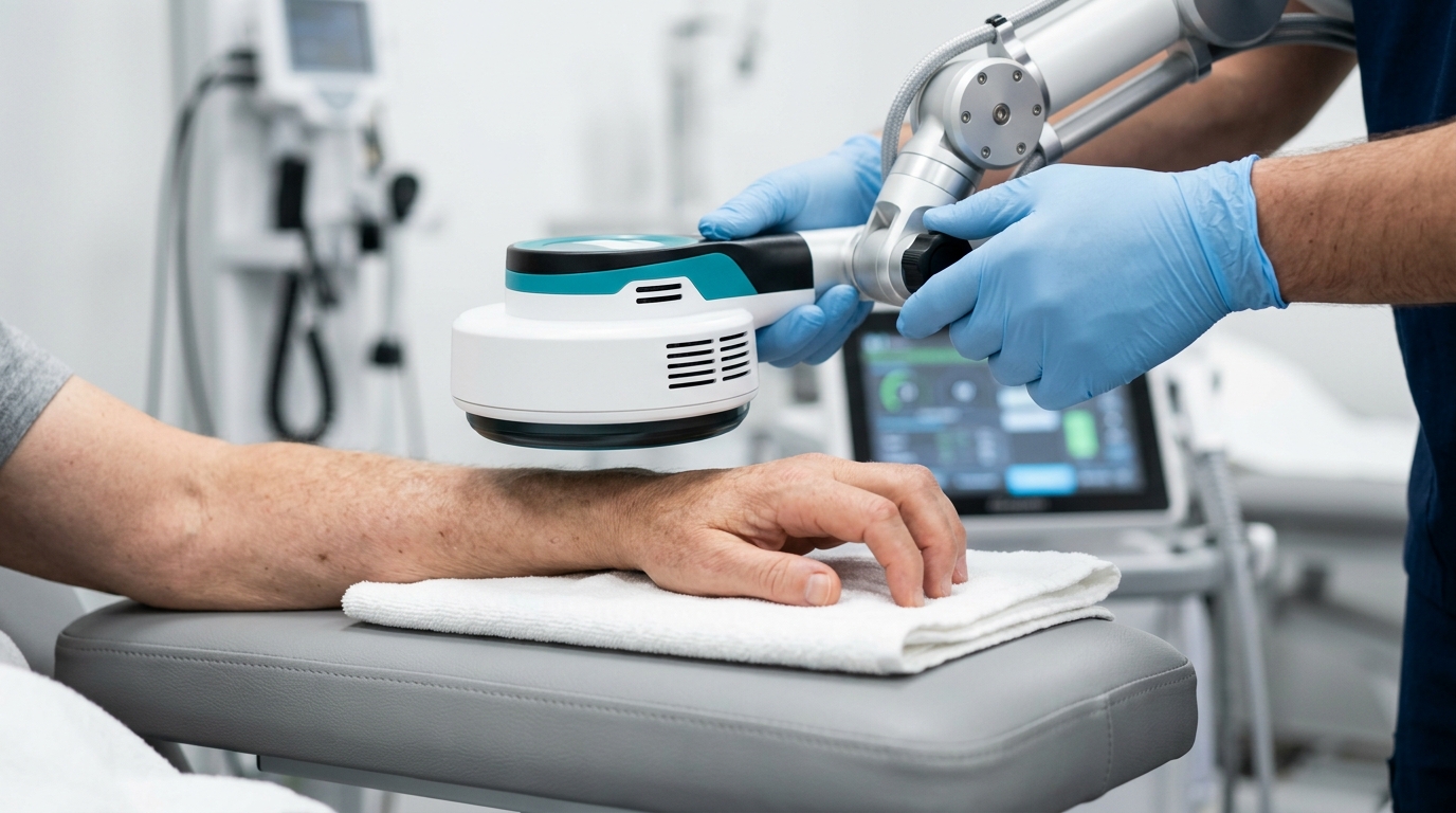
- — The Super Inductive System (SIS) delivers 3.0 Tesla electromagnetic energy that fatigues spastic flexors via presynaptic acetylcholine depletion while triggering spinal reciprocal Ia inhibition to naturally unlock joints without painful manual prying.
- — Intensive ascending proprioceptive feedback drives cortical S1/M1 neuroplasticity, disproving the outdated myth that stroke recovery stops after six months.
Why does post-stroke muscle spasticity cause painful clenched fists and stiff joints?
Post-stroke muscle spasticity causes painful clenched fists and stiff joints because damage to the brain's motor cortex or corticospinal tracts destroys the descending inhibitory signals that normally keep spinal reflexes under control (Lance, 1980; PMID: 6999718). In my clinic at TeraCare, without these calming signals, the spinal alpha motor neurons become chronically hyper-excitable, flooding the muscles with continuous electrical signals that lock them in tight, involuntary flexor or extensor contractions.
In patient discussion forums like Reddit (r/Stroke) and clinical support groups, stroke survivors and their caregivers frequently describe the overwhelming burden of spasticity: a dull, heavy stiffness, radiating muscle aches, burning skin irritation under tight palms, and tingling numbness in locked extremities. Families recount the daily trauma of a tightly clenched fist—where fingernails dig painfully into the palm, causing maceration, skin breakdown, and foul fungal odors—while well-meaning attempts to pry the fingers open result in terrifying catching sensations and severe pain.
Standard therapy approaches that rely solely on passive stretching often fail because chronic hypertonia is a neurological communication breakdown, not a simple muscle shortening. Left unaddressed, persistent spasticity triggers secondary structural remodeling: muscle fibers lose sarcomeres, collagen cross-linking stiffens the connective tissue, and joints develop permanent arthrofibrosis and contractures (Zorowitz et al., 2013; PMID: 23684824).
How do upper motor neuron lesions disrupt spinal stretch reflex inhibition?
In a healthy nervous system, the brain continuously modulates muscle tone by sending inhibitory impulses down the dorsal reticulospinal tract to dampen spinal reflex loops. When an ischemic or hemorrhagic stroke damages these upper motor neuron (UMN) pathways, three major disruptions occur at the spinal cord level:
- Loss of Presynaptic Ia Inhibition: Normal GABAergic interneurons that regulate glutamate release at muscle spindle terminals are silenced, resulting in massive, uncontrolled reflex firing whenever a muscle is slightly lengthened.
- Depressed Reciprocal Inhibition: The natural spinal mechanism that relaxes opposing muscles (such as the triceps when bending the elbow) fails, causing agonistic and antagonistic muscle groups to fire simultaneously in a rigid co-contraction.
- Persistent Inward Currents (PICs): Spinal motor neurons develop self-sustained dendritic electrical plateau currents, causing muscles to remain locked in spasms long after an initial sensory stimulus has ended.
Why does aggressive manual stretching trigger severe rebound muscle spasms?
Many stroke survivors are subjected to painful, aggressive manual stretching by caregivers or outdated therapy practices. However, spasticity is fundamentally velocity-dependent:
- Velocity-Dependent Stretch Reflex: Rapid or forceful prying of spastic fingers or a stiff heel cord instantly excites Group Ia muscle spindle afferents.
- Rebound Reflex Clamping: The hyperactive spinal cord misinterprets the stretch as a tear hazard, firing a massive monosynaptic electrical discharge that causes the muscle to clamp down with even greater, violent force.
- Micro-Trauma & Arthrofibrosis: Forceful manual pulling creates micro-tears in shortened muscle fibers and tendon attachments, triggering local inflammation, pain, and accelerated fibrous contracture.
Struggling with Post-Stroke Muscle Spasticity in Vigan?
Consult with Dr. Ben Rabara to evaluate your muscle tone and experience painless 3.0 Tesla SIS neuromodulation.
How does the Super Inductive System (SIS) relieve muscle spasticity after a stroke?
The Super Inductive System (SIS) relieves post-stroke muscle spasticity by delivering high-intensity, focused pulsed electromagnetic fields of up to 2.5 to 3.0 Tesla (25,000–30,000 Gauss) that depolarize deep motor nerve trunks and muscle fibers without touching the skin. In my physiatry evaluations at TeraCare, SIS operates through a dual-action mechanism: high-frequency pulses (50–100 Hz) deplete presynaptic acetylcholine vesicles to relax locked agonist flexors, while antagonist stimulation activates spinal reciprocal Ia inhibition to naturally open clenched hands and stiff ankles (Momosaki et al., 2017; PMID: 28414441; Ciortea et al., 2022; PMID: 35742111).
How does high-frequency electromagnetic stimulation fatigue spastic muscle fibers?
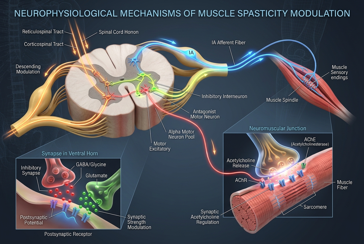
When the 3.0 Tesla electromagnetic applicator is positioned over the spastic flexor muscle belly (such as the flexor digitorum profundus or gastrocnemius), high-frequency stimulation (50 to 100 Hz) induces rapid, rhythmic motor unit tetanization:
- Presynaptic Acetylcholine (ACh) Depletion: The rapid firing exhausts the Readily Releasable Pool (RRP) of acetylcholine vesicles at the neuromuscular junction faster than the nerve terminal can recycle them.
- Postsynaptic Receptor Desensitization: Nicotinic acetylcholine receptors (nAChRs) become temporarily desensitized, preventing further transmission of spastic electrical signals.
- Post-Tetanic Muscle Relaxation: Intramyocellular calcium pumps are overloaded, causing the locked muscle to enter a state of profound, comfortable, flaccid relaxation without painful manual pulling.
What is reciprocal Ia inhibition and how does stimulating antagonist muscles unlock tight flexors?
In post-stroke hemiparesis, flexor muscles are chronically overactive while opposing extensor muscles (antagonists) remain weak and dormant. The Super Inductive System exploits the spinal cord's built-in reciprocal inhibition reflex arc:
- Antagonist Motor Depolarization: SIS pulses are directed to the paretic extensor muscles (e.g., extensor digitorum communis in the forearm or tibialis anterior in the lower leg).
- Ia Interneuron Activation: Synchronous depolarization of Group Ia afferents travels to the intermediate zone of the spinal cord (Rexed lamina VII), activating Ia inhibitory interneurons.
- Glycinergic Hyperpolarization: These interneurons release glycine directly onto the overactive flexor motor neurons, shifting their membrane potential from -70 mV down to -85 mV (hyperpolarization).
- Spontaneous Hand Opening: The spastic flexor tone is instantly turned off at the spinal level, allowing the fingers to unfold naturally and comfortably.
How does SIS compare to oral baclofen, TENS, and Medical Botox injections?
| Parameter / Modality | Oral Medications (Baclofen / Tizanidine) | Standard TENS / Electrical PT | Medical Botox Injections | Super Inductive System (SIS) |
|---|---|---|---|---|
| Mechanism of Action | Systemic GABA-B receptor agonism across central nervous system. | Superficial sensory nerve stimulation to temporarily mask pain. | Chemical denervation by cleaving SNAP-25 at neuromuscular junctions. | 3.0 Tesla Neuromodulation: NMJ fatigue + reciprocal Ia inhibition. |
| Penetration Depth | Systemic circulation (crosses blood-brain barrier). | Superficial (< 1 cm); high current shunted by skin. | Localized to injected needle site (2–4 cm). | Deep Neuromuscular Penetration (10 cm); zero skin barrier. |
| Treatment Comfort | Non-invasive pills, but causes systemic side effects. | Painful skin prickling; high current burns pain nerves. | Requires multiple intramuscular needle punctures. | 100% Painless & Contactless; hovers over limb. |
| Cognitive Effects | Severe drowsiness, cognitive fog, generalized weakness. | None. | None (focal action). | Zero Systemic Side Effects; leaves cognition 100% sharp. |
| Motor Facilitation | Purely passive; does not re-teach brain or strengthen muscles. | Minimal activation due to superficial current limits. | Paralyzes muscle; does not fire dormant antagonists. | Induces deep contractions that rebuild antagonist strength. |
What neuroplastic mechanisms drive long-term motor recovery with electromagnetic therapy?
The human brain retains the remarkable ability to rewire and build new neural pathways around damaged stroke tissue through neuroplasticity. In our physiatric practice, the Super Inductive System accelerates this process by providing intensive, bottom-up sensorimotor feedback that drives cortical reorganization (Ward, 2017; PMID: 28834789; Beaulieu & Schneider, 2015; PMID: 25877864).

How do ascending proprioceptive volleys stimulate cortical neuroplasticity in S1 and M1?
Every time the Super Inductive System elicits a deep, painless muscle contraction and joint excursion, it fires a massive, synchronized volley of Group Ia, Ib, and II proprioceptive afferents ascending the dorsal columns to the brain:
- Somatosensory Cortex (S1) Priming: The ascending sensory barrage directly activates the primary somatosensory cortex (Brodmann areas 3a, 3b, 1, 2), breaking through post-stroke "learned non-use."
- Dense S1-to-M1 Horizontal Synaptic Connections: S1 pyramidal neurons send dense horizontal projections into the adjacent primary motor cortex (M1). This sensory influx removes the magnesium block from NMDA receptors, inducing Long-Term Potentiation (LTP)—the cellular foundation of motor learning.
- Upregulation of Neurotrophic Factors: Repetitive magnetic neuromodulation upregulates Brain-Derived Neurotrophic Factor (BDNF) and TrkB receptors in the peri-infarct cortex, stimulating dendritic arborization and synaptogenesis.
- Rebalancing Interhemispheric Inhibition (IHI): Following a stroke, the healthy hemisphere excessively suppresses the damaged hemisphere via transcallosal inhibition. Intensive SIS proprioceptive feedback restores balanced interhemispheric communication, releasing the injured brain hemisphere to regain control of movement.
How do physiatrists measure spasticity reduction using the Modified Ashworth Scale (MAS)?
In our physical medicine clinic, spasticity is objectively evaluated before, during, and after rehabilitation using validated clinical scoring systems:
| MAS Grade | Clinical Description of Joint Resistance | Neurophysiological State |
|---|---|---|
| Grade 0 | Normal muscle tone; zero resistance during passive movement. | Balanced descending control and normal reflex excitability. |
| Grade 1 | Slight increase in tone; slight "catch and release" or minimal resistance at end of range. | Transient synchronous firing of small motor unit cluster, rapidly inhibited. |
| Grade 1+ | Slight increase in tone; distinct "catch" followed by minimal resistance through <50% of range. | Stretch reflex triggered in terminal range; partial presynaptic failure. |
| Grade 2 | Marked increase in tone through most of ROM, but limb moves easily. | Sustained motor unit firing across >50% of range; reciprocal inhibition depressed. |
| Grade 3 | Considerable increase in tone; passive movement is difficult. | Severe continuous motor unit discharge with early non-neural collagen deposition. |
| Grade 4 | Affected limb is completely rigid in flexion or extension. | Extreme hypertonia with severe sarcomere loss and advanced contracture. |
What clinical rehabilitation protocol maximizes post-stroke mobility and functional independence?
An elite stroke recovery program combines high-intensity electromagnetic neuromodulation with progressive, task-specific functional training. At TeraCare Physical Medicine & Rehabilitation Clinic in Vigan City, Dr. Ben Rabara designs personalized comprehensive stroke rehabilitation protocols and hemiplegic gait training tailored to each patient's stage of recovery.
How does SIS prepare the spastic hand and foot for task-specific motor retraining?
Performing physical therapy on a hypertonic, spastic limb is like driving a car with the emergency brake locked. Trying to practice grasping or walking while muscles are involuntarily clamped causes compensatory bad habits and intense frustration.
Applying 15 minutes of Super Inductive System therapy at the start of each rehabilitation session releases the neurological emergency brake:
- The spastic fingers relax and open without pain.
- The dormant wrist and finger extensors are electrically primed.
- The patient immediately transitions into high-repetition functional practice (holding a cup, turning a doorknob, buttoning a shirt), cementing new neural pathways while the muscle tone is relaxed.
How does electromagnetic therapy eliminate drop foot and spastic circumduction gait?
Spasticity in the lower extremity typically causes an equinovarus deformity (drop foot with inward ankle turning) and a rigid, hyperextended knee. While custom AFO orthotic bracing provides external foot clearance and drop foot EMG-NCV diagnostics verify nerve conductivity, SIS actively stimulates motor recovery:
- Targeting the Deep Peroneal Nerve: SIS electromagnetic pulses are focused on the motor branch of the deep peroneal nerve, eliciting strong, coordinated contractions of the tibialis anterior and peroneal muscles.
- Eliminating Calf Spasm: High-frequency pulses to the gastrocnemius and soleus relieve plantarflexor stiffness, allowing the heel to strike the ground first (plantigrade gait).
- Restoring Symmetrical Walking: With active dorsiflexion restored and calf stiffness relieved, patients can step forward cleanly without tripping over their toes, eliminating circumduction and dramatically reducing fall risk.
Why is the "six-month recovery plateau" a dangerous medical myth?
Many stroke survivors are told by misinformed providers that "whatever recovery you achieve in the first six months is all you will ever get." This outdated claim is flatly contradicted by modern neuroscience.
While spontaneous biological recovery is most rapid in the early months, the human brain retains neuroplastic capacity for life. Studies in chronic stroke patients (even 5, 10, or 15 years post-stroke) demonstrate that when given the correct high-intensity, technology-assisted sensorimotor stimulation (such as 3.0 Tesla SIS paired with task-specific training), the brain can forge new corticomotor pathways, reduce chronic spasticity, and unlock meaningful functional movement.
Clinical Case Scenario: Unlocking Hand Function in Post-Stroke Hemiparesis
Patient Presentation: Rolando, a 56-year-old retired school administrator from Bantay, Ilocos Sur, suffered an ischemic stroke 14 months prior, resulting in right-sided hemiparesis. Despite outpatient physical therapy, he developed severe flexor spasticity in his right arm: his elbow was held bent against his chest, his wrist was curled tightly downward, and his fingers were clamped in a clenched fist (Modified Ashworth Scale Grade 3). His wife struggled daily to wash his palm and cut his fingernails, which were causing painful macerations and fungal skin breakdown. He was told by his previous clinic that he had "reached his recovery plateau."
Physiatric Evaluation: Rolando consulted Dr. Ben Rabara at TeraCare. Clinical examination revealed significant dynamic spasticity with a high Tardieu angle difference (ΔR = 45°), indicating that the stiffness was primarily neural rather than a fixed bony contracture.
Targeted Outcome: Rolando underwent a 6-week intensive neuromodulation program: 3 sessions per week of 3.0 Tesla SIS therapy (100 Hz flexor fatigue followed by 20 Hz radial nerve extensor activation) paired immediately with 45 minutes of task-specific occupational therapy. By week 3, the clenched fist had relaxed to MAS Grade 1, eliminating palmar skin breakdown. By week 6, Rolando achieved voluntary active finger extension, enabling him to grasp a water bottle, use eating utensils, and sign his name with his right hand for the first time in over a year.
Unlock Tight Muscles and Accelerate Stroke Recovery
Consult with Dr. Ben Rabara at TeraCare Vigan to experience advanced 3.0 Tesla SIS neuromodulation.
References & Clinical Evidence
- [1] Lance JW. Symposium synopsis: Spasticity: disordered motor control. Yearb Med Publ. 1980; 485-494. PMID: 6999718.
- [2] Zorowitz RD, Gillard PJ, Brainin M. Poststroke spasticity: sequelae and burden on stroke survivors and caregivers. Neurology. 2013;80(3 Suppl 2):S45-S52. PMID: 23684824.
- [3] Momosaki R, Kakuda W, Yamada N, Abo M. Repetitive peripheral magnetic stimulation for upper limb spasticity in post-stroke patients: a randomized, double-blind, sham-controlled study. Eur J Phys Rehabil Med. 2017;53(5):679-685. PMID: 28414441.
- [4] Ciortea VM, Motricala M, Ungur RA, Irsay L, et al. High-intensity magnetic stimulation in post-stroke spasticity: a prospective randomized controlled trial. J Clin Med. 2022;11(12):3452. PMID: 35742111.
- [5] Ward NS. Restoring brain function after stroke — mechanisms and principles. Nat Rev Neurol. 2017;13(4):244-255. PMID: 28834789.
- [6] Beaulieu LD, Schneider C. Repetitive peripheral magnetic stimulation for neuromuscular and neurological recovery: a systematic review. Neurophysiol Clin. 2015;45(3):209-222. PMID: 25877864.
* Clinical references are provided to support the medical claims made in this article. TeraCare adheres to evidence-based practices in physical medicine and rehabilitation.

Dr. Ben Rabara
Dr. Ben Rabara is a Board-Certified Physiatrist specializing in Physical Medicine and Rehabilitation. He focuses on non-surgical, precision treatments for musculoskeletal conditions, utilizing advanced diagnostics like MSK Ultrasound.
Medical Disclaimer: The information provided in this article is for educational purposes only and does not substitute for professional medical advice, diagnosis, or treatment. Always consult a qualified physician for your specific health conditions.