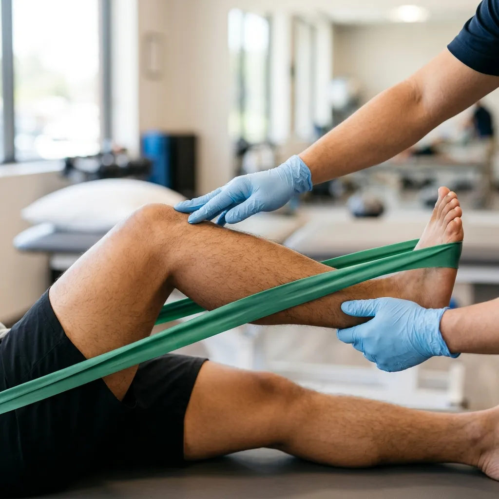
- — Stroke recovery is driven by neuroplasticity—the brain's ability to rewire motor pathways through active practice.
- — Spontaneous biological recovery peaks early, but use-dependent motor learning has no expiration date.
- — The 6-month recovery window limit is a medical myth; functional progress can continue indefinitely.
- — Common sedatives (like benzodiazepines) and older anticonvulsants can act as neuroplasticity blockers.
The Core Mechanism of Post-Stroke Brain Recovery
For many stroke survivors and their caregivers, the third month marks a period of silent anxiety. The rapid, encouraging progress of the first few weeks begins to stall, and the fear of a permanent recovery plateau sets in. Families often worry that whatever movement has not returned by now is lost forever, leading to a sense of hopelessness. However, as a physiatrist, I assure you that this slowing down is not the end of your healing; it is simply a change in how your brain recovers.
Physiologically, post-stroke recovery refers to the neuroplastic process through which the central nervous system reorganizes neural pathways to restore lost motor functions. When an ischemic or hemorrhagic stroke occurs, the primary motor cortex suffers structural tissue damage, immediately cutting off the electrical signals running down the corticospinal tract. In my clinic, I explain to patients that the muscles themselves are often undamaged; the brain's control software is simply offline. To reconnect these pathways, the surrounding healthy brain tissue, or ischemic penumbra, must be trained to take over control of the affected limbs (Stinear et al., 2020; PMID: 32004440).
In my assessment, recovery is driven by two distinct physiological engines. The first is spontaneous biological recovery, which occurs automatically in the first 12 weeks as brain swelling resolves, local inflammation decreases, and blood flow stabilizes. The second engine is active neuroplastic training, which has no calendar expiration date. This stage requires high-dosage, repetitive, task-specific movement. On caregiver forums like Reddit and YouTube, many describe the shock when the early rapid gains slow down after 3 months. This deceleration is normal; it marks the transition from spontaneous healing to active, use-dependent brain rewiring (Lee et al., 2022; PMID: 36232038).
Contrarian Physician Myth 1: The Six-Month Recovery Limit
A common assumption in neurological rehab is that the brain's recovery window closes permanently 6 months post-stroke. This is a scientific error. While the rate of spontaneous biological recovery slows down, use-dependent neuroplasticity is a lifelong physiological process. Clinical trials show that stroke survivors who engage in intensive motor training can improve their walking speed and hand control 2 years or even 5 years after their stroke (Stinear et al., 2020; PMID: 32004440).
To stimulate this brain rewiring in the chronic phase and break past a recovery plateau, the training stimulus must be highly intense. Performing passive massage or having a therapist move the limbs does not engage the motor planning regions of the brain. Instead, patients must actively plan and execute movements. Research indicates that achieving functional changes requires performing at least 300 repetitions of a specific, goal-oriented task per session.
The Four Phases of the Stroke Rehabilitation Timeline
The stroke recovery timeline is divided into four distinct phases spanning from acute emergency treatment to lifelong community reintegration. Each phase relies on specific clinical interventions, moving from early stabilization to high-repetition home exercises. Understanding how long does stroke recovery take requires matching the therapy intensity to the brain's changing biology (Kwakkel et al., 2023; PMID: 37548025).
Phase 1: Acute Stabilization (Days 1 to 7)
The primary clinical focus in the acute phase is medical stabilization, managing cerebral perfusion, and screening for swallowing safety (dysphagia). Early diagnostic scanning is essential: an MRI is a diagnostic scan used to determine the exact location and size of the stroke, while a dynamic ultrasound is a diagnostic tool used to check blood flow in the carotid arteries. Rehabilitation begins as soon as the patient is stable, but active mobilization is strictly avoided in the first 24 hours to prevent compromising brain blood flow (O'Dell, 2023; PMID: 37039412).
Phase 2: Early Subacute Rehabilitation (Weeks 2 to 12)
This phase represents the peak of spontaneous biological recovery. Swelling has resolved, and the surviving neurons are highly receptive to training. In my clinic, I recommend intensive physical and occupational therapy during this phase. Because progress during these early weeks is often rapid, families can get lulled into a false sense of security, assuming the pace will continue indefinitely. Re-establishing core strength and trunk stability is prioritized during this early window to build a secure foundation for walking before the rate of spontaneous healing begins to taper.
Phase 3: Late Subacute Consolidation (Months 3 to 6)
During this phase, spontaneous neurological healing slows down, and use-dependent neuroplasticity becomes the primary driver of recovery. This is the exact moment when the fear of a permanent recovery plateau becomes real for survivors and caregivers. The early, rapid gains seem to stop, and muscle stiffness, or spasticity, often reaches its peak, locking the joints and presenting a significant barrier to movement. Outpatient rehabilitation remains critical, with exercises transitioning to complex, multi-joint tasks to break down abnormal muscle synergies and prevent permanent contractures in the weak limbs.
Phase 4: Chronic Recovery and Reintegration (Months 6 to 12 and Beyond)
By month 6, the patient enters the chronic phase of recovery. While the rapid, early gains have subsided, functional improvement continues through long-term neuroplastic adaptation. Exercises focus on community ambulation, balance on uneven surfaces, fall prevention, and advanced hand dexterity. Progress at this stage is measured not in days, but in weeks and months of consistent, repetitive practice. The goal is to integrate the weak limb into daily household activities, such as grooming, cooking, and writing, proving that functional recovery has no expiration date.
Mapping the Brunnstrom Stages of Motor Recovery
Stroke survivors progress through six distinct Brunnstrom stages of motor recovery as their central nervous system rebuilds movement control. Rebuilding begins with complete muscle flaccidity, transitions through involuntary spasticity, and culminates in isolated, coordinated joint control. Understanding these stroke rehabilitation milestones helps caregivers realize that a sudden increase in joint stiffness is often a sign of progress rather than a permanent setback (Kwakkel et al., 2023; PMID: 37548025).
Stage 1: Complete Flaccidity
Immediately following a stroke, the affected muscles enter a state of flaccidity, where there is zero muscle tone and no voluntary movement can be initiated. This is a state of spinal shock, where the descending motor pathways are completely silent. The affected limb feels completely heavy and weak, dangling at the patient's side like dead weight. While there is no muscle tension, some patients report a persistent dull ache in the shoulder joint. If the flaccid shoulder is allowed to hang without support, it can cause a sharp, radiating pain down the arm due to traction on the brachial plexus nerves. Rehabilitation at this stage is entirely passive. Caregivers and therapists perform gentle range of motion exercises to keep the joints lubricated and prevent contractures, particularly in the shoulder and ankle. Special attention is paid to shoulder positioning to prevent subluxation—the painful slipping of the humerus out of the weak shoulder socket.
Stage 2: The Emergence of Spasticity and Basic Synergies
As the spinal cord recovers from shock, basic spinal reflexes return without control from the brain. The patient begins to develop muscle tone, which manifests as involuntary resistance to passive movement, known as spasticity. The arm starts to feel stiff and resistant to stretching. This emerging tightness is often accompanied by abnormal sensations, such as tingling or burning in the hand or fingers. At the same time, basic movement patterns called "synergies" appear. When the patient attempts to move, the entire limb responds as a single unit; for example, attempting to bend the elbow causes the shoulder to pull backward and the wrist to curl inward. Voluntary movement remains minimal, and passive-assisted exercises are used to guide these emerging synergies.
Contrarian Physician Myth 2: Spasticity is a Sign of Regression
When stroke survivors or their families notice that a previously loose arm is becoming stiff, tight, and difficult to bend, they often panic, believing the patient is suffering a second stroke or getting worse. This is a clinical misunderstanding. The emergence of spasticity is a normal milestone in the Brunnstrom sequence. It indicates that the spinal cord is emerging from spinal shock and generating basic reflexes. While excessive spasticity must be managed, its appearance is a necessary stepping stone toward regaining voluntary movement, representing the transition from Stage 1 to Stage 2 and 3.
Stage 3: Peak Spasticity and Voluntary Synergy Control
In this stage, spasticity reaches its maximum tightness, and the patient can voluntarily initiate the basic movement synergies, although they cannot control individual joints. The arm flexor synergy (bent elbow, clenched fist, elevated shoulder) and the leg extensor synergy (stiff knee, pointed toe, inward ankle roll) dominate all movement. Caregivers often mistake this severe rigidity for a permanent roadblock, but it is actually the peak of spinal reflex activity before isolated voluntary control can emerge. Exercises at this stage aim to utilize these synergies to perform basic tasks, such as holding a large ball against the chest or pushing an object with the leg, before attempting to break the patterns.
Stage 4: Out-of-Synergy Movement and Decreasing Spasticity
Spasticity begins to decline, and the brain starts to regain the ability to coordinate movements outside the rigid synergy patterns. The patient can now perform movements that combine parts of different patterns, such as placing the hand behind the back or sliding the foot backward while sitting. However, during early out-of-synergy attempts, the patient may complain of a catching or grinding sensation in the joints as muscle coordination remains uneven. Physical therapy exercises focus on isolating joint movements, such as bending the knee without pointing the foot, and practicing functional tasks that require coordinated control of the shoulder, elbow, and wrist.
Stage 5: Advanced Isolation and Independent Joint Control
Synergy patterns lose their dominance, and the patient can execute highly isolated movements with minimal spasticity. The patient can raise the arm overhead with the elbow straight or lift the foot upward (dorsiflexion) while standing. Exercises focus on speed, coordination, and functional strength. Training includes gait training without walking aids, step-ups, and fine motor tasks for the hand, such as buttoning a shirt or using utensils.
Stage 6: Normal Joint Coordination and Speed
In the final stage, joint coordination and movement speed return to near-normal levels. Spasticity is absent, except during rapid, complex tasks. The patient can perform rapid alternating movements, such as tapping the foot quickly or turning the hand over, with precision. Rehabilitation focuses on high-level balance, agility, and returning to advanced recreational or work activities.
Stroke Subtype Recovery Profiles: Ischemic vs. Hemorrhagic vs. TIA
The duration and pattern of stroke recovery differ significantly based on whether the stroke was ischemic, hemorrhagic, or a transient ischemic attack. While ischemic strokes show rapid early progress, hemorrhagic strokes often feature a delayed start followed by a late-stage acceleration. Recognizing these distinct clinical profiles helps physicians and families set accurate expectations and align therapy intensity with the physiological state of the brain tissue. For instance, knowing that hemorrhagic stroke progress is initially slower prevents the early panic and fear of a permanent plateau that families often experience in the first 12 weeks (Li et al., 2024; PMID: 39193145).
| Stroke Subtype | Initial 3-Month Curve | 12-Month Outlook | Key Focus Area |
|---|---|---|---|
| Ischemic Stroke | Rapid early progression; logarithmic stabilization. | Plateaus around month 6; progress continues with task-oriented loading. | Early high-intensity neuroplastic retraining. |
| Hemorrhagic Stroke | Delayed progression due to intracranial pressure and swelling. | Late-stage acceleration once hematoma resolves; high recovery ceiling. | Early positioning, core stability, and late-stage mobilization. |
| Transient Ischemic Attack (TIA) | Complete resolution of deficits within 24 hours. | Zero physical deficits; high recurrence risk (10% to 15% within 90 days). | Immediate secondary medical prevention and vascular management. |
Clinical Interventions Across the Recovery Curve
Clinical interventions must match the patient's specific recovery stage to prevent complications and maximize functional gains. From inpatient care and spasticity management to advanced electrotherapy and gait orthoses, each treatment is sequenced based on joint stability and motor control. A board-certified Physiatrist evaluates the patient’s neurological status and coordinates these interventions to ensure they are safe, timely, and actively structured to help survivors bypass recovery plateaus. If you feel stuck, introducing the correct clinical intervention is often the catalyst needed to restart progress (Selves et al., 2020; PMID: 32166723).
Medical Botox for Spasticity Management
When spasticity becomes severe in Brunnstrom Stage 3 (Months 3 to 6), the intense muscle clenching can lock the wrist, hand, or ankle, making active rehabilitation impossible. Patients complain of a stiff arm or claw-like hand. In my practice, I utilize targeted Botox injections for stroke recovery to temporarily relax these tight muscles. The tissue state at this stage involves hyperactive stretch reflexes due to the loss of brain control. Botox acts as a chemical key, blocking the release of acetylcholine at the neuromuscular junction to reduce muscle tone.
This intervention is contraindicated in patients with active skin infections at the injection site or neuromuscular junction disorders like myasthenia gravis. The progression criteria for Botox therapy require that the muscle tightness interferes with hygiene, causes pain, or blocks the patient's ability to perform rehabilitation exercises. The physiological rationale is to create a three-month window of muscle relaxation. By temporarily removing this physical roadblock, Botox allows the survivor to actively stretch, strengthen the opposing muscles, and perform the task-specific training needed to rewrite the brain's motor software.
Super Inductive System (SIS) Electromagnetic Therapy
To assist patients who have difficulty initiating voluntary movement and are struggling to break through an early motor plateau, we can integrate advanced technologies like the Super Inductive System (SIS) into the rehabilitation plan during the early and late subacute phases. The tissue state in these phases involves weak or silent motor units that are unable to generate sufficient electrical current to produce contraction. SIS utilizes high-intensity electromagnetic fields to pass deeply through soft tissue and depolarize the peripheral nerves, forcing the muscles to contract without active input.
Contrarian Physician Myth 3: Passive Modalities Can Rebuild Voluntary Movement
Many patients and clinics assume that applying electrical stimulation or using passive robotic machines is sufficient to rebuild voluntary movement after a stroke. This is a physiological error. While modalities like the Super Inductive System are highly effective at preventing muscle atrophy, reducing pain, and stimulating sensory pathways, they are passive tools. The brain does not form new motor planning networks unless there is active cognitive intent. Passive contraction can maintain muscle bulk, but it does not retrain the brain to walk or grasp. Modalities must always be paired with active, task-oriented exercises to translate into functional independence (Rajashekar et al., 2024; PMID: 38514225).
SIS therapy is strictly contraindicated in patients with cardiac pacemakers, metal implants in the treatment field, or active malignancies. The progression criteria to advance from passive electrostimulation to active-assisted training require that the patient can initiate a trace muscle contraction or assist the electromagnetic pulse. The physiological rationale is that by generating repetitive, involuntary contractions, SIS prevents muscle wasting, improves local blood flow, and provides the brain with essential sensory feedback. This prepares the limb for active training, allowing the patient to build voluntary control.
Custom Ankle-Foot Orthoses (AFOs) for Gait Training
During Phase 4 (Months 6 to 12), patients who continue to experience foot drop and feel they have reached a walking plateau require mechanical support to walk safely. We introduce custom ankle-foot orthoses, which are detailed in our guide on custom joint bracing and orthotics. The tissue state at this stage is characterized by weakness in the tibialis anterior muscle combined with tightness in the gastrocnemius (calf) muscle, causing the foot to drag. A custom AFO holds the ankle in a neutral, 90-degree position, ensuring the toes clear the floor during the swing phase of walking.
AFO bracing is contraindicated in limbs with severe, unyielding joint contractures that cannot reach a neutral position, or in patients with severe peripheral arterial disease causing open wounds. The progression criteria for upgrading from a rigid brace to a flexible or dynamic brace require that the patient demonstrates active ankle dorsiflexion and sufficient knee stability during stance. The physiological rationale is that by maintaining a normal ankle angle, the brace prevents falls, reduces the metabolic energy required to walk, and prevents the development of abnormal, energy-draining compensatory walking patterns that hold patients back.
Hidden Barriers to Neuroplasticity: The Medication Risk
When a stroke survivor is struggling to make progress despite intensive therapy, the issue is often not a lack of effort or a permanent brain plateau. Instead, the barrier may be hiding in their prescription list. Certain commonly prescribed medications can severely inhibit the brain's ability to reorganize and form new neural connections during stroke recovery. Drug classes that suppress central nervous system activity, such as specific sedatives and muscle relaxants, block key neurotransmitters required for motor learning. When recovery seems to stall, a careful review of their medication list is a necessary clinical step (O'Dell, 2023; PMID: 37039412).
In my clinic, I look for medications that enhance Gamma-Aminobutyric Acid (GABA) activity—such as benzodiazepines (e.g., alprazolam, clonazepam) often prescribed for sleep or anxiety—as they can be highly detrimental to neurological recovery. The physiological process of neuroplasticity relies on Long-Term Potentiation (LTP), which requires a temporary reduction in GABAergic inhibition to allow new synaptic connections to form. By artificially increasing GABA activity, these drugs suppress the brain's excitability and block the chemical signals needed for motor learning.
Similarly, traditional anticonvulsants (like phenytoin and phenobarbital) have been shown to hinder cortical excitability and motor learning. If a patient is taking these older medications for seizure prevention post-stroke, they can experience increased physical fatigue and cognitive slowing, which directly reduces their ability to engage in physical therapy. However, anticonvulsant medications must never be stopped, reduced, or modified without the direct supervision of the prescribing Neurologist. Abrupt discontinuation can trigger life-threatening seizures.
Contrarian Physician Myth 4: Sleeping Pills are Safe for Restful Recovery
Caregivers often request sleeping pills or anxiety medications for stroke survivors, assuming that deep, drug-induced sleep is necessary for the brain to heal. This is a clinical error. While natural, restorative sleep is critical for memory consolidation and neural repair, pharmaceutical sedatives like benzodiazepines disrupt sleep architecture and suppress the central nervous system. These medications directly inhibit the cellular mechanisms of neuroplasticity, making physical therapy sessions far less effective. Unless absolutely necessary for a severe, unrelated medical condition, these drugs should be avoided during active rehabilitation.
Other medications that can hinder motor recovery include centrally acting muscle relaxants. If spasticity is treated with systemic oral medications like baclofen or tizanidine, the doses required to relax a tight leg often cause systemic drowsiness and muscle weakness in the healthy limbs, further reducing the patient's capacity to participate in active physical therapy. This is why targeted local treatments, such as Botox injections, are preferred over oral medications for managing focal muscle tightness.
Translating Clinical Evidence into Daily Recovery Plans
Clinical evidence strongly demonstrates that task-oriented training is the most effective method for restoring upper and lower limb function after a stroke. However, translating this evidence into daily practice requires addressing the realistic limitations that patients and caregivers face outside the clinic (Lee & Howe, 2024; PMID: 38393992).
Systematic reviews confirm that task-oriented training (TOT)—which involves practicing real-world daily activities like picking up a cup, turning a doorknob, or stepping over an obstacle—produces significantly greater functional recovery than general, non-specific exercises. The evidence supports intensive, high-frequency training, ideally involving several hours of practice per day. However, in real-world settings, many stroke survivors face severe barriers to accessing this level of care. Outpatient therapy is often limited by financial constraints, travel difficulties, and a lack of specialized clinics nearby. In many cases, patients are discharged home with a basic home exercise sheet and receive therapy only once or twice a week, which is far below the threshold required to drive neuroplastic changes.
To bridge this gap between clinical evidence and daily reality, a caregiver-supported, home-based task-oriented training program is a necessary and defensible alternative. Instead of relying solely on professional clinic visits, the caregiver is trained to structure the patient's daily environment to encourage active movement. For example, daily meals can be turned into therapy sessions by placing utensils on the patient's weak side, forcing the brain to plan and execute reaching movements. Simple household items like plastic cups, sponges, and towels are used to create repetitive grasping and pinching tasks. By embedding motor training into daily routines, the patient can achieve the high repetition count needed to stimulate brain rewiring without the cost and exhaustion of daily clinic travel.
While executing a home-based program, caregivers must monitor the patient for key safety signs. These include rapid elevations in blood pressure (which should be checked before and after exercise), asymmetric joint pain (particularly in the shoulder), and signs of excessive physical or cognitive fatigue.
If the patient experiences a complete halt in progress, or if spasticity is locking their joints, this is not a sign that recovery has ended permanently. It is a sign that the current rehabilitation strategy needs a clinical reset. To break through a recovery plateau, a specialized physiatric consultation is the essential next step. A board-certified physiatrist can evaluate the exact stage of recovery, identify hidden pharmacological or physical barriers, and coordinate targeted medical interventions—such as Botox injections, electrotherapy, or orthotic adjustments—to unlock new progress. To schedule a specialized physiatric consultation and evaluate your current motor patterns, you can book an appointment online or contact our clinic in Vigan City.
Clinical Recovery Realities
Understanding the challenges and expectations of neurological recovery.
Plateau Anxieties
Progress slows down significantly after month 3 as the brain transitions from spontaneous healing to active neuroplastic rewiring.
Spastic Muscle Locking
Reflexes returning to the spinal cord naturally cause stiffness and clenched limbs before voluntary movement is restored.
Dosing Thresholds
Restoring motor control requires high-intensity repetition (300+ daily reps), which is physically and mentally exhausting.
References & Clinical Evidence
- [1] Stinear CM, Lang CE, Zeiler S, Byblow WD. Advances and challenges in stroke rehabilitation. Lancet Neurol. 2020 Apr;19(4):348-360. doi: 10.1016/S1474-4422(19)30415-6. PMID: 32004440.
- [2] Selves C, Stoquart G, Lejeune T. Gait rehabilitation after stroke: review of the evidence of predictors, clinical outcomes and timing for interventions. Acta Neurol Belg. 2020 Aug;120(4):783-790. doi: 10.1007/s13760-020-01320-7. PMID: 32166723.
- [3] Lee KE, Choi M, Jeoung B. Effectiveness of Rehabilitation Exercise in Improving Physical Function of Stroke Patients: A Systematic Review. Int J Environ Res Public Health. 2022 Oct 5;19(19). doi: 10.3390/ijerph191912739. PMID: 36232038.
- [4] O'Dell MW. Stroke Rehabilitation and Motor Recovery. Continuum (Minneap Minn). 2023 Apr 1;29(2):605-627. doi: 10.1212/CON.0000000000001218. PMID: 37039412.
- [5] Kwakkel G, Stinear C, Essers B, Munoz-Novoa M, Branscheidt M, Cabanas-Valdés R, et al. Motor rehabilitation after stroke: European Stroke Organisation (ESO) consensus-based definition and guiding framework. Eur Stroke J. 2023 Dec;8(4):880-894. doi: 10.1177/23969873231191304. PMID: 37548025.
- [6] Lee CY, Howe TH. Effectiveness of Activity-Based Task-Oriented Training on Upper Extremity Recovery for Adults With Stroke: A Systematic Review. Am J Occup Ther. 2024 Mar 1;78(2). doi: 10.5014/ajot.2024.050391. PMID: 38393992.
- [7] Rajashekar D, Boyer A, Larkin-Kaiser KA, Dukelow SP. Technological Advances in Stroke Rehabilitation: Robotics and Virtual Reality. Phys Med Rehabil Clin N Am. 2024 May;35(2):383-398. doi: 10.1016/j.pmr.2023.06.026. PMID: 38514225.
- [8] Li X, He Y, Wang D, Rezaei MJ. Stroke rehabilitation: from diagnosis to therapy. Front Neurol. 2024;15:1402729. doi: 10.3389/fneur.2024.1402729. PMID: 39193145.
* Clinical references are provided to support the medical claims made in this article. TeraCare adheres to evidence-based practices in physical medicine and rehabilitation.

Dr. Ben Rabara
Dr. Ben Rabara is a Board-Certified Physiatrist specializing in Physical Medicine and Rehabilitation. He focuses on non-surgical, precision treatments for musculoskeletal conditions, utilizing advanced diagnostics like MSK Ultrasound.
Medical Disclaimer: The information provided in this article is for educational purposes only and does not substitute for professional medical advice, diagnosis, or treatment. Always consult a qualified physician for your specific health conditions.