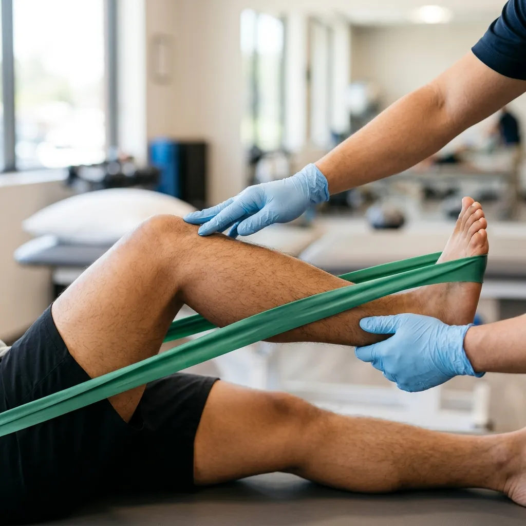
- — Nonpharmacological interventions, particularly active exercise and stabilization, are the recommended first-line treatment for low back pain and sciatica.
- — Absolute bed rest impairs spinal function; gentle, guided movement is required to maintain core strength and disc health.
- — Sciatica recovery relies on directional preference, utilizing extension drills to migrate herniated disc gel away from compressed nerve roots.
- — Deep core stabilization exercises like McGill's McGill Big Three or DNS training build deep endurance without loading the joints.
Slipped Disc vs. Muscle Strain: How a Physiatrist Differentiates Back Pain
Differentiating a herniated disc from a muscular strain requires a detailed clinical examination of neurological indicators and movement triggers. While a muscle strain presents with localized tenderness and pain during stretching, a herniated disc exhibits dermatomal numbness, diminished reflexes, and a positive Straight Leg Raise test. Diagnostic accuracy ensures correct treatment selection (Chou, 2021; PMID: 34370518).
When back pain strikes, patients often struggle to understand if they have simply pulled a muscle or suffered a structural disc injury ("slipped disc"). A muscular strain involves microscopic tears in the erector spinae or quadratus lumborum muscles, usually caused by sudden lifting or awkward twisting. While painful and tender to touch, muscle strains do not compromise the nervous system. The pain remains localized to the lower back, does not radiate below the knee, and typically resolves within a few weeks as the tissue heals.
Conversely, a herniated disc occurs when the tough outer ring of the intervertebral disc (annulus fibrosus) tears, allowing the soft inner gel (nucleus pulposus) to bulge outward. This gel can mechanically compress or chemically irritate the adjacent spinal nerve roots (most commonly the L4, L5, or S1 levels). This nerve root compression is what produces true radiculopathy, or sciatica—a sharp, electric shock-like pain that travels down the buttock, thigh, and outer calf, sometimes reaching the foot. A physiatrist confirms this diagnosis through specific neurological testing: assessing dermatomal sensation, measuring deep tendon reflexes, and performing the Straight Leg Raise (Lasègue's sign), which stretches the inflamed nerve root over the herniated disc material to reproduce leg symptoms between 30 and 70 degrees.
Contrarian Physician Myth 1: The MRI Myth
A common patient belief is that an MRI is required immediately to identify the cause of back pain, and that any bulge found is a structural disaster requiring surgery. This is clinically incorrect. MRI findings must be correlated with a physical examination, as disc bulges and degeneration are highly prevalent in asymptomatic populations (Han et al., 2023; PMID: 37096189). Over 30% of healthy adults in their 30s with zero pain have herniated discs on imaging (Øiestad et al., 2020; PMID: 31433541). We treat the patient's functional deficits, not the scan.
Many online portals advocate for immediate, aggressive spinal stretches, such as forward folds, to relieve lower back tightness. In a patient with a herniated disc, bending forward increases the mechanical load on the anterior portion of the lumbar vertebrae. This compressive force pushes the herniated nucleus pulposus further posterior, increasing nerve root compression and worsening sciatica. Standard forward-bending stretches must be strictly avoided during acute herniation phases.
Relieving Sciatica: How Directional Preference Relieves Pinched Nerves
Sciatica relief depends on identifying the patient's directional preference to centralize radiating nerve pain. By utilizing McKenzie extension protocols (such as prone press-ups), the physical therapist applies mechanical pressure to migrate the herniated nucleus pulposus anteriorly. This movement offloads the compressed L4-S1 nerve roots, reducing peripheral leg symptoms (Karlsson et al., 2020; PMID: 32795336).
The cornerstone of mechanical diagnosis and therapy (MDT) for disc herniations is the "centralization" phenomenon. Centralization occurs when repeated movements shift the radiating pain from the foot or calf back up toward the lower back. This movement indicates that the mechanical compression on the nerve root is decreasing. Extension is the most common directional preference for posterior disc herniations, as it uses the natural lordotic curve of the lumbar spine to push the herniated gel forward, away from the nerve.
Sciatica Extension Protocol
For extension-tolerant patients, we establish the following progressive protocol:
- Tissue State: A posterior or posterior-lateral disc protrusion compressing the S1 or L5 nerve root, with symptoms intolerant to lumbar flexion.
- Contraindication Logic: McKenzie extension exercises are contraindicated if the patient has spinal stenosis with severe claudication, or if extension causes the pain to travel further down the leg (peripheralization).
- Prone Lying (Step 1): The patient lies flat on their stomach on a firm surface. If the pain is highly irritable, place a pillow under the pelvis. Rest for 2–3 minutes.
- Prone on Elbows (Step 2): Prop up onto the elbows, keeping the lower back and hips completely relaxed, letting the pelvis sag into the mattress. Breathe deeply for 1–2 minutes.
- Prone Press-Ups (Step 3): Place hands flat under the shoulders and slowly push the chest upward, straightening the elbows. The hips and lower back must remain completely passive. Perform 10 slow repetitions, breathing out at the top, every 2 hours while awake.
- Progression Criteria: We progress the patient from prone press-ups to standing lumbar extension only when they can achieve full passive extension in prone without reproducing radiating leg pain.
- Physiological Rationale: Lumbar extension compresses the posterior aspect of the intervertebral disc. This mechanical wedge action encourages the displaced nucleus pulposus to migrate anteriorly, reducing mechanical tension and chemical irritation on the adjacent nerve root.
Stabilizing the Spine: Core Exercises for Herniated Discs
Spine stabilization exercises build deep core endurance to protect a herniated disc from unstable shear forces. Rather than performing lumbar flexion movements, patients train spinal stiffness using McGill's stabilization exercises (the bird-dog, side plank, and modified curl-up). This clinical progression builds muscular support around the lumbar spine without causing structural compression (Huang et al., 2025; PMID: 40025463).
A herniated disc leaves the motion segment of the spine micro-instable. To protect the healing tissue, the surrounding muscles must act as a stabilizer. However, traditional "abdominal strength" exercises, like sit-ups and crunches, are highly destructive for herniated discs. Performing a crunch forces the lumbar spine into flexion under load, which places massive compression on the disc and pushes the nucleus pulposus backward. Spine rehabilitation should instead focus on building isometric endurance to prevent movement of the lumbar spine during activity.
Dynamic Neuromuscular Stabilization (DNS) training activates deep stabilizer networks, including the transversus abdominis (TrA) and the diaphragm (Huang et al., 2025; PMID: 40025463). In the clinic, we use Rehabilitative Ultrasound Imaging (RUSI) as visual biofeedback to help patients see their deep muscles contracting in real-time, which has been shown to be superior to verbal or tactile cues for muscle recruitment (Valera-Calero et al., 2021; PMID: 34300002).
Contrarian Physician Myth 2: The Core Strength Myth
Many patients assume that building a strong core requires training the visible "six-pack" rectus abdominis muscle. For back pain rehabilitation, rectus abdominis strength is largely irrelevant. What matters is the endurance and coordinated contraction of the deep stabilizers—the transversus abdominis, lumbar multifidus, and pelvic floor. These muscles act as a natural weightlifting belt to stiffen the spine and prevent painful micro-movements.
Many online workout routines recommend dynamic lumbar twisting (Russian twists) or deep toe touches to "stretch" a sore back. From a biomechanical perspective, combining lumbar flexion with rotation is the exact mechanism that ruptures the fibers of the annulus fibrosus, causing a disc herniation. Forcing a herniated disc through twisting under load is a primary cause of acute flare-ups and progressive nerve compression.
The Limitations of Passive Care: Why Active Exercise Beats Ultrasound
Passive modalities, such as therapeutic ultrasound, provide short-term sensory relief but fail to correct the underlying biomechanical instability of chronic low back pain. Cochrane systematic reviews show no clinical justification for ultrasound in disc rehabilitation. Long-term recovery requires active motor control retraining to restore structural load-bearing capacity and pelvic stability (Ebadi et al., 2020; PMID: 32623724).
It is common for clinics to rely heavily on passive modalities, such as hot packs, electrical stimulation (TENS), traction, and therapeutic ultrasound, to treat back pain. While these treatments are comforting and can temporarily reduce pain signaling (gating mechanism), they do not change the structural load tolerance of the spine. A Cochrane Database Systematic Review analyzed 10 randomized trials and concluded that the evidence does not support the use of therapeutic ultrasound in the management of chronic low back pain (Ebadi et al., 2020; PMID: 32623724).
Active exercise remains the gold standard for long-term functional recovery (Flynn, 2020; PMID: 33064421). Rather than laying passively on a treatment table, patients must engage in structured movement to stimulate tissue remodeling (mechanotransduction) and rebuild the neuromuscular control of the pelvis and trunk. Passive care should only be used as a brief preparatory window to lower pain levels enough to allow active training to begin.
Contrarian Physician Myth 3: The Heat and Ice Cure-All
Many believe that applying heat or ice is a sufficient treatment to cure back pain. While ice vasoconstricts vessels to temporarily numb acute pain, it reduces the delivery of healing factors. Heat increases local blood flow but does not build muscle endurance or stabilize a herniated disc. Relying on temperature patches ignores the mechanical dysfunction driving the pain; they are comfort measures, not cures.
Anatomical Mimics: Is it Really Sciatica?
Shooting leg pain does not always originate from a herniated disc; it may be referred from sacroiliac joint injury or piriformis muscle entrapment. Differentiating these mimics requires orthopedic provocation tests, sensory mapping, and nerve conduction studies. Identifying the correct culprit ensures that physical therapy exercises target the true biomechanical driver of pain (Stretanski et al., 2026; PMID: 32491804).
"Sciatica" is a symptom, not a diagnosis. Any condition that compresses or irritates the sciatic nerve or its roots can produce radiating leg pain. As a result, there are several anatomical mimics that present similarly to a herniated disc:
- Sacroiliac Joint (SIJ) Dysfunction: The SIJ connects the pelvis to the spine, absorbing shear forces during walking. Irritation of this joint can refer pain down the posterior thigh, mimicking L5-S1 disc pain. However, SIJ injury is characterized by localized joint tenderness, a positive pain provocation cluster (distraction, compression, Gaenslen's tests), and the absence of midline back pain (Stretanski et al., 2026; PMID: 32491804).
- Lumbar Facet Joint Arthropathy: The small joints connecting the vertebrae can develop osteoarthritis. Bending backward or twisting loads these joints, referring pain into the gluteal region. A physiatrist separates facet pain from disc pain because facet pain is worsened by spinal extension (Kemp's test), while disc herniations are typically intolerant to spinal flexion.
- Piriformis Syndrome: The sciatic nerve passes directly under or through the piriformis muscle in the buttock. If this muscle becomes tight or spasmed, it can mechanically entrap the nerve. This condition is differentiated by deep buttock tenderness and pain during the FAIR test (flexion, adduction, internal rotation), with no neurological deficits (like reflex loss) in the lower leg.
Standard online advice frequently conflates hamstring tightness with sciatic nerve irritation, recommending deep hamstring stretches (like forward bending). For a patient with true discogenic sciatica, pulling on the hamstring places the sciatic nerve under severe neural tension, stretching the already inflamed nerve root over the herniated disc and significantly worsening the radiculopathy. Hamstring stretching should be avoided during acute sciatica flares.
Evidence-to-Practice: Reconciling Guidelines with Local Reality
Clinical practice guidelines prioritize active exercise and cognitive-behavioral pacing as first-line therapies for low back pain, reserving surgery and MRIs for progressive motor deficits. In provincial settings like Vigan, implementing this requires adapting high-cost clinical setups into structured, home-based stabilization programs. This practical approach ensures rehabilitation consistency while reducing out-of-pocket costs (Chou, 2021; PMID: 34370518).
The 2021 American College of Physicians guidelines emphasize that nonpharmacological exercise and cognitive pacing should be the first-line therapies for low back pain, reserving expensive imaging (like MRIs) for cases with progressive neurological loss (Chou, 2021; PMID: 34370518). In an ideal clinical system, back pain patients attend supervised, multidisciplinary rehabilitation 3 times a week, utilizing real-time ultrasound feedback (RUSI) to verify multifidus recruitment.
However, in provincial settings like Vigan, Ilocos Sur, patients face travel constraints, farming work schedules, and high out-of-pocket costs that make frequent clinic visits impossible. To translate this evidence into practice, we conduct an in-clinic diagnostic assessment to rule out red flags, identify the patient's directional preference, and teach deep core activation. We then transition the patient to a structured home program (using a calendar checklist to track adherence), where they perform 3 sets of McGill's modified curl-up and bird-dog daily, returning to the clinic only once every two weeks for progression checks. We monitor for progressive leg weakness or bowel/bladder changes, escalating to urgent specialist care or imaging if red flags emerge.
Dr. Rabara's Spine Rehabilitation Clinic in Vigan
Spine rehabilitation under Dr. Ben Paolo C. Rabara at TeraCare Clinic in Vigan combines physiatric diagnostic screening, EMG-guided nerve studies, and personalized physical therapy blueprints. By coordinating active exercise with spasticity management, dynamic ultrasound imaging, and patient education, we help back pain survivors regain mechanical stability.
Every spine is unique, and generic exercises can easily worsen a herniated disc if directional preference is ignored. At TeraCare Clinic in Vigan, Dr. Rabara performs a comprehensive physiatric assessment, mapping lumbar mechanical triggers, sensory loss, and joint range of motion. We design individualized movement blueprints executed under the close supervision of certified physical therapists.
We actively coordinate care with local families, adapting rehabilitation plans to accommodate Northern Luzon transit realities and out-of-pocket cost constraints, offering options for structured home programs to ensure progress continues between clinic visits.
Rebuild Your Spinal Blueprint Safely
Recovering from a herniated disc or sciatica requires a structured, medically supervised pathway. A physiatric movement assessment helps identify your specific directional preference, screen for joint contractures, and establish a safe, progressive rehabilitation plan tailored to your lifestyle and resources.
Schedule a Spine EvaluationWritten by: Dr. Ben Paolo C. Rabara
Specialty: Physical Medicine & Rehabilitation (Physiatry)
Location: Vigan, Ilocos Sur, Philippines
Last reviewed: 2026-06-19
Disclaimer: This page is for educational and informational purposes only and does not constitute medical advice. Sciatica and low back pain can be signs of serious neurological compression, and recovery requires an individualized assessment and guidance from a licensed physician.
References & Clinical Evidence
- [1] Chou R. Low Back Pain. Ann Intern Med. 2021;174(8):ITC113-ITC128. PMID: 34370518.
- [2] Ebadi S, Henschke N, Forogh B, Nakhostin Ansari N, et al. Therapeutic ultrasound for chronic low back pain. Cochrane Database Syst Rev. 2020;7(7):CD009169. PMID: 32623724.
- [3] Huang H, Xie H, Zhang G, Xiao W, et al. Effects of Dynamic Neuromuscular Stabilization on chronic low back pain. BMC Musculoskelet Disord. 2025;26(1):112. PMID: 40025463.
- [4] Han CS, Hancock MJ, Sharma S, Sharma S, et al. Low back pain of disc, sacroiliac joint, or facet joint origin: a diagnostic accuracy systematic review. EClinicalMedicine. 2023;59:101960. PMID: 37096189.
- [5] Stretanski MF, Gutcho J, Hanna A. Sacroiliac Joint Injury. StatPearls. 2026. PMID: 32491804.
- [6] Valera-Calero JA, Fernández-de-Las-Peñas C, Varol U, Ortega-Santiago R, et al. Ultrasound Imaging as a Visual Biofeedback Tool in Rehabilitation: An Updated Systematic Review. Int J Environ Res Public Health. 2021;18(14):7554. PMID: 34300002.
* Clinical references are provided to support the medical claims made in this article. TeraCare adheres to evidence-based practices in physical medicine and rehabilitation.

Dr. Ben Rabara
Dr. Ben Rabara is a Board-Certified Physiatrist specializing in Physical Medicine and Rehabilitation. He focuses on non-surgical, precision treatments for musculoskeletal conditions, utilizing advanced diagnostics like MSK Ultrasound.
Medical Disclaimer: The information provided in this article is for educational purposes only and does not substitute for professional medical advice, diagnosis, or treatment. Always consult a qualified physician for your specific health conditions.