- — Stroke recovery relies on neuroplasticity, which is the brain's ability to reorganize and form new neural connections through repetitive, structured movement.
- — Passive therapies alone cannot restore motor patterns; active, task-specific exercises are essential for rebuilding walking confidence and leg control.
- — Foot drop rehabilitation requires targeted dorsiflexion exercises to retrain the tibialis anterior muscle and prevent gait imbalances.
- — Physiatric supervision ensures that home programs are adapted for safety, addressing local socioeconomic barriers and preventing fall injuries.
Neuroplasticity: Re-educating the Brain's Movement Software
Neuroplasticity serves as the primary mechanism for neurological recovery, allowing the central nervous system to reorganize neural pathways after a stroke. By performing repetitive, active-oriented physical exercises, stroke survivors stimulate axonal sprouting and synaptic remodeling around the damaged cortical zones. This structured input helps the brain rewrite its motor control software to restore voluntary limb control (GBD 2021 Stroke Risk Factor Collaborators, 2024; PMID: 39304265).
When an ischemic or hemorrhagic stroke occurs, the primary motor cortex suffers tissue damage, which immediately disrupts the descending signals running through the corticospinal tract. This injury manifests as hemiparesis—the loss of voluntary motor control on one side of the body. In the clinic, we explain to patients that the muscles themselves are often undamaged; rather, the "electrical wiring" and the brain's control programs have been offline. To reconnect these pathways, we cannot rely on passive modalities. Passive stretching or having a caregiver move the leg provides essential sensory feedback, but it does not engage the frontal lobe's motor planning centers.
Without active cognitive effort and physical practice, patients fall into a state of "learned non-use," where the brain permanently deletes the representation of the paretic limb from its cortical map. Studies confirm that chronic stroke sensorimotor impairment is associated with secondary structural changes in the brain, including smaller hippocampal volumes (Zavaliangos-Petropulu et al., 2022; PMID: 35574963). This secondary degeneration highlights the critical necessity of active, cognitive-motor training. Engaging in high-repetition, task-specific training (such as grasping a cup or stepping over a line) forces the brain to recruit adjacent, undamaged cortical areas to assume control of the weak muscles.
Contrarian Physician Myth 1: The Bed Rest Myth
A common assumption among patients and family members is that absolute bed rest is the safest response after a stroke to prevent a recurrence. However, complete immobilization is clinically counterproductive. Prolonged bed rest induces rapid skeletal muscle atrophy, losing up to 5% of muscle mass per week of immobilization, while severely reducing proprioceptive input to the brain, which reinforces learned non-use. Unless the patient exhibits hemodynamic instability, active rehabilitation should begin as soon as cleared by the attending physician.
Many online articles claim that passive massage is a sufficient substitute for physical therapy. In clinical reality, while passive stroking temporarily increases local blood flow, it fails to engage the motor planning regions of the frontal lobe. Without active muscular contraction and cognitive intent, the brain cannot form new synaptic connections. Over-reliance on passive stretching or rubbing can actually delay the recovery window, leaving the patient with permanent motor deficits that could have been mitigated by active motor re-education.
Rebuilding Foot Control: Exercises for Stroke-Induced Drop Foot
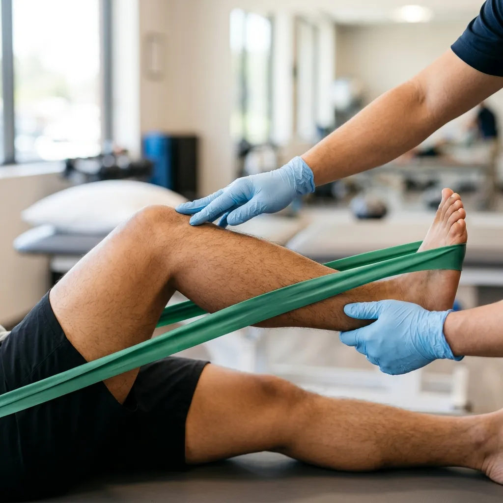
Drop foot exercises target the tibialis anterior muscle to restore ankle dorsiflexion and prevent tripping during the swing phase of walking. Repetitive, active-assisted ankle movements, such as towel curls and band-resisted dorsiflexion, promote cortical reorganization. These clinical movements retrain the deep peroneal nerve pathway, preventing the foot from dragging and improving safety while walking (Roots et al., 2022; PMID: 35380302).
Healthy walking requires at least 10 degrees of ankle dorsiflexion (lifting the toes upward) to clear the ground during the swing phase of gait. When spasticity or paralysis affects the tibialis anterior, the foot hangs downward, causing the toes to catch on the floor. To compensate, stroke survivors often adopt abnormal walking patterns, such as "circumduction gait" (swinging the weak leg outward in a semi-circle) or "steppage gait" (lifting the hip abnormally high). These compensations increase the metabolic cost of walking by over 50%, leading to early fatigue and placing severe mechanical strain on the lumbar spine and pelvis.
Drop Foot Clinical Protocol
To target this specific motor deficit, we implement a structured rehabilitation protocol:
- Tissue State Assessment: We evaluate the degree of voluntary control over the ankle dorsiflexors and measure the passive joint range. Post-stroke muscle stiffness in the medial gastrocnemius (calf muscle) often acts as a physical barrier, which can be measured quantitatively using ultrasound elastography (Roots et al., 2022; PMID: 35380302).
- Contraindication Logic: Active dorsiflexion exercises are modified or delayed if the patient has a fixed ankle contracture, severe joint instability, or a suspected deep vein thrombosis (DVT) in the paretic calf.
- Active-Assisted Dorsiflexion: The patient sits in a chair, loops a strap or towel around the ball of the paretic foot, and holds the ends. As they attempt to actively pull their toes toward their shin, they use their hands to pull the strap, providing active assistance at the end of the range. Perform 3 sets of 10 repetitions, twice daily.
- Progression Criteria: We progress the patient to active-free dorsiflexion (lifting the foot without the strap) once they can achieve 5 degrees of active dorsiflexion without ankle inversion (turning the foot inward). Resisted training using light elastic bands is introduced only when active range of motion is stable.
- Physiological Rationale: Repetitive ankle movement drives mechanotransduction in the muscle spindle fibers of the tibialis anterior. This sensory feedback travels up the deep peroneal nerve to the motor cortex, stimulating synaptic plasticity and helping the brain rebuild voluntary control over the foot.
Retraining the Sit-to-Stand: Restoring Functional Leg Strength
Sit-to-stand training after a stroke rebuilds quadriceps strength and motor control needed for daily independence. The exercise involves leaning slightly forward, pushing evenly through both feet, and standing before slowly returning to a seated position. Using a high, sturdy chair minimizes knee shear and builds confidence before progressing to lower surfaces (Li H, et al., 2026; PMID: 41581201).
The ability to rise from a chair is a critical functional benchmark. However, hemiparetic patients naturally develop a habit of shifting 80% of their body weight onto their healthy leg when standing up. This asymmetry deprives the paretic leg of the mechanical loading needed to rebuild quadriceps and gluteal strength, while causing pelvic tilt and chronic hip pain over time. Retraining this movement requires breaking it down into three distinct motor phases:
- Flexion Momentum Phase: The patient slides their hips forward to the edge of the chair, places their feet slightly behind the knees, and leans the trunk forward ("nose over toes") to shift their center of mass.
- Momentum Transfer Phase: The patient pushes down through both feet, activating the gluteus maximus and quadriceps to lift the hips off the seat.
- Extension Phase: The patient straightens their hips and knees to achieve upright standing, ensuring equal weight distribution across both legs.
Contrarian Physician Myth 2: The Symmetrical Standing Myth
A common mistake is instructing stroke survivors to stand up without correcting their foot placement, assuming that "any stand is a good stand." This reinforces hemiparetic neglect. To force neuromuscular re-education, we must place the healthy foot slightly forward (mechanically unloading it) or place a small wooden block under the healthy foot. This arrangement forces the brain to load and activate the weak leg to complete the stand.
The 2024 Lancet Neurology study emphasizes that stroke is a leading cause of long-term disability, and early rehabilitation is vital to restore functional independence (GBD 2021 Stroke Risk Factor Collaborators, 2024; PMID: 39304265). In an ideal clinical setting, post-stroke sit-to-stand training is performed under constant supervision using specialized harness systems and body-weight support treadmills. However, in provincial settings like Vigan, Ilocos Sur, patients face travel barriers and the financial burden of attending multiple weekly sessions out-of-pocket. To translate this evidence into practice, we instruct family members to configure a high dining chair (at least 45 centimeters tall) against a wall and assist the patient in performing 3 sets of 8 repetitions daily. The caregiver stands on the affected side to block the knee from buckling, providing a safe, zero-cost home adaptation of the clinical protocol. We monitor for sudden gait asymmetry or fatigue, escalating back to in-clinic assessment if the patient experiences new joint pain or cannot rise safely after three weeks.
Understanding Spasticity: Managing Muscle Stiffness After Stroke
Post-stroke muscle spasticity arises from hyperactive stretch reflexes due to upper motor neuron damage. Managing this stiffness requires combining daily passive stretching with active-assisted range of motion exercises to prevent structural muscle contractures. This clinical approach reduces tone, maintains joint alignment, and prepares the limb for active strengthening under physiatric guidance (Roots et al., 2022; PMID: 35380302).
Spasticity is not simple muscle tightness; it is a neurological condition where the stretch reflex remains hyperactive because the damaged brain can no longer send inhibitory "calm down" signals. As a result, even a minor movement causes the muscle to contract involuntarily, leading to painful stiffness, clenched fists, or a foot pulled downward and inward (equinovarus deformity). If left untreated, this constant contraction leads to structural changes in the muscle fibers, which lose their elasticity and shorten permanently, forming a joint contracture.
Quantitative research using ultrasound elastography confirms that stroke survivors experience significantly increased muscle stiffness in the paretic limbs compared to healthy sides (Roots et al., 2022; PMID: 35380302). To combat this stiffness, we recommend a daily passive stretching protocol: the caregiver slowly moves the joint to its end-range (e.g., pulling the foot upward) and holds the stretch for 30 seconds, repeating 3 to 5 times. The stretch must be slow; a rapid stretch will trigger the hyperactive reflex and worsen the stiffness.
Contrarian Physician Myth 3: The Stretching Fixes Everything Myth
Many believe that daily stretching alone is enough to cure spasticity and restore movement. This is false. Passive stretching only provides a short-term reduction in muscle stiffness (lasting from minutes to a few hours) and does not retrain the brain. Stretching should be used as a preparatory window: we stretch the stiff muscle to restore range of motion, and immediately follow it with active exercises to strengthen the opposing muscle group, teaching the brain how to actively control the new range.
Typical online guides recommend generic balance exercises like standing on one foot or walking on foam. For a stroke survivor, this is dangerous and biomechanically inappropriate. A paretic ankle has impaired reflex responses; forcing a patient onto unstable surfaces without correcting their gait symmetry triggers high-amplitude compensatory movements, worsening their limp and increasing fall risk. Balance training must start with static weight-shifting on stable, hard ground, slowly introducing dynamic steps only when the patient has achieved pelvic stability during stance phase.
Relearning Balance: Exercises to Prevent Falls After Stroke
Balance retraining after a stroke begins by restoring proprioceptive awareness and core strength. By practicing supported standing drills against a wall or using parallel bars, the patient stimulates mechanoreceptors in the feet. This clinical progression allows the nervous system to coordinate trunk muscles, reducing fall risks without compromising joint alignment (Li H, et al., 2026; PMID: 41581201).
Following a stroke, the brain struggles to process where the body is in space (proprioception). This sensory deficit, combined with weakness in the core and gluteal muscles, makes stroke survivors feel highly unstable, leading to a profound fear of falling. Re-waking these balance networks requires a progressive, safe exercise sequence:
- Supported Wall Standing: The patient stands with their back and hips flat against a wall, feet hip-width apart. This setup provides a safe tactile reference, helping the brain \"feel\" vertical alignment and reducing the fear of falling backward.
- Weight-Shifting Drills: Standing facing a sturdy table or countertop, holding on with both hands, the patient slowly shifts their weight onto the paretic leg, holds for 5 seconds, and shifts back. This stimulates the mechanoreceptors in the sole of the foot, sending crucial sensory signals back to the brain. Perform 10 shifts per side, 3 times daily.
- Step-Up Exercises: Once weight-shifting is stable, the patient practices stepping forward with the healthy leg while keeping the weak leg flat on the ground. This drill forces the paretic leg to act as a stable anchor, rebuilding stance-phase hip and knee control.
Dr. Rabara's Neurological Rehabilitation Clinic in Vigan
Stroke rehabilitation under Dr. Ben Paolo C. Rabara at TeraCare Clinic in Vigan combines physiatric diagnostic screening, EMG-guided nerve studies, and personalized physical therapy blueprints. By coordinating active exercise with spasticity management (such as medical Botox or bracing), we help stroke survivors transition safely from clinical dependence to functional community mobility.
Every stroke survivor presents a unique clinical picture, depending on the brain lesion's size and location. A generic list of home exercises found online cannot account for spasticity levels, cardiac risks, or joint contractures. At TeraCare Clinic in Vigan, Dr. Rabara performs a comprehensive physiatric assessment, mapping motor deficits, sensory loss, and joint range of motion. We design individualized movement blueprints executed under the close supervision of certified physical therapists.
We actively coordinate care with local families, adapting rehabilitation plans to accommodate Northern Luzon transit realities and out-of-pocket cost constraints, offering options for structured home programs to ensure progress continues between clinic visits.
Rebuild Your Movement Blueprint Safely
Recovering from a stroke requires a structured, medically supervised pathway. A physiatric movement assessment helps identify your specific motor deficits, screen for joint contractures, and establish a safe, progressive rehabilitation plan tailored to your lifestyle and resources.
Schedule a Rehabilitation EvaluationWritten by: Dr. Ben Paolo C. Rabara
Specialty: Physical Medicine & Rehabilitation (Physiatry)
Location: Vigan, Ilocos Sur, Philippines
Last reviewed: 2026-06-19
Disclaimer: This page is for educational and informational purposes only and does not constitute medical advice. A stroke is a complex neurological emergency and recovery requires an individualized assessment and guidance from a licensed physician.
References & Clinical Evidence
- [1] GBD 2021 Stroke Risk Factor Collaborators. Global, regional, and national burden of stroke and its risk factors, 1990-2021: a systematic analysis for the Global Burden of Disease Study 2021. Lancet Neurol. 2024;23(10):973-1003. PMID: 39304265.
- [2] Roots J, Trajano GS, Fontanarosa D. Ultrasound elastography in the assessment of post-stroke muscle stiffness: a systematic review. Insights Imaging. 2022;13(1):52. PMID: 35380302.
- [3] Zavaliangos-Petropulu A, Lo B, Donnelly MR, Schweighofer N, et al. Chronic Stroke Sensorimotor Impairment Is Related to Smaller Hippocampal Volumes: An ENIGMA Analysis. J Am Heart Assoc. 2022;11(10):e025109. PMID: 35574963.
- [4] Li H, Zhao Z, Qiao P, Wang J, et al. Meta-Analysis of Treatment Methods for Poststroke Cognitive Impairment: A Network Analysis of Various Interventions. Brain Behav. 2026;16(2):e371115. PMID: 41581201.
* Clinical references are provided to support the medical claims made in this article. TeraCare adheres to evidence-based practices in physical medicine and rehabilitation.

Dr. Ben Rabara
Dr. Ben Rabara is a Board-Certified Physiatrist specializing in Physical Medicine and Rehabilitation. He focuses on non-surgical, precision treatments for musculoskeletal conditions, utilizing advanced diagnostics like MSK Ultrasound.
Medical Disclaimer: The information provided in this article is for educational purposes only and does not substitute for professional medical advice, diagnosis, or treatment. Always consult a qualified physician for your specific health conditions.