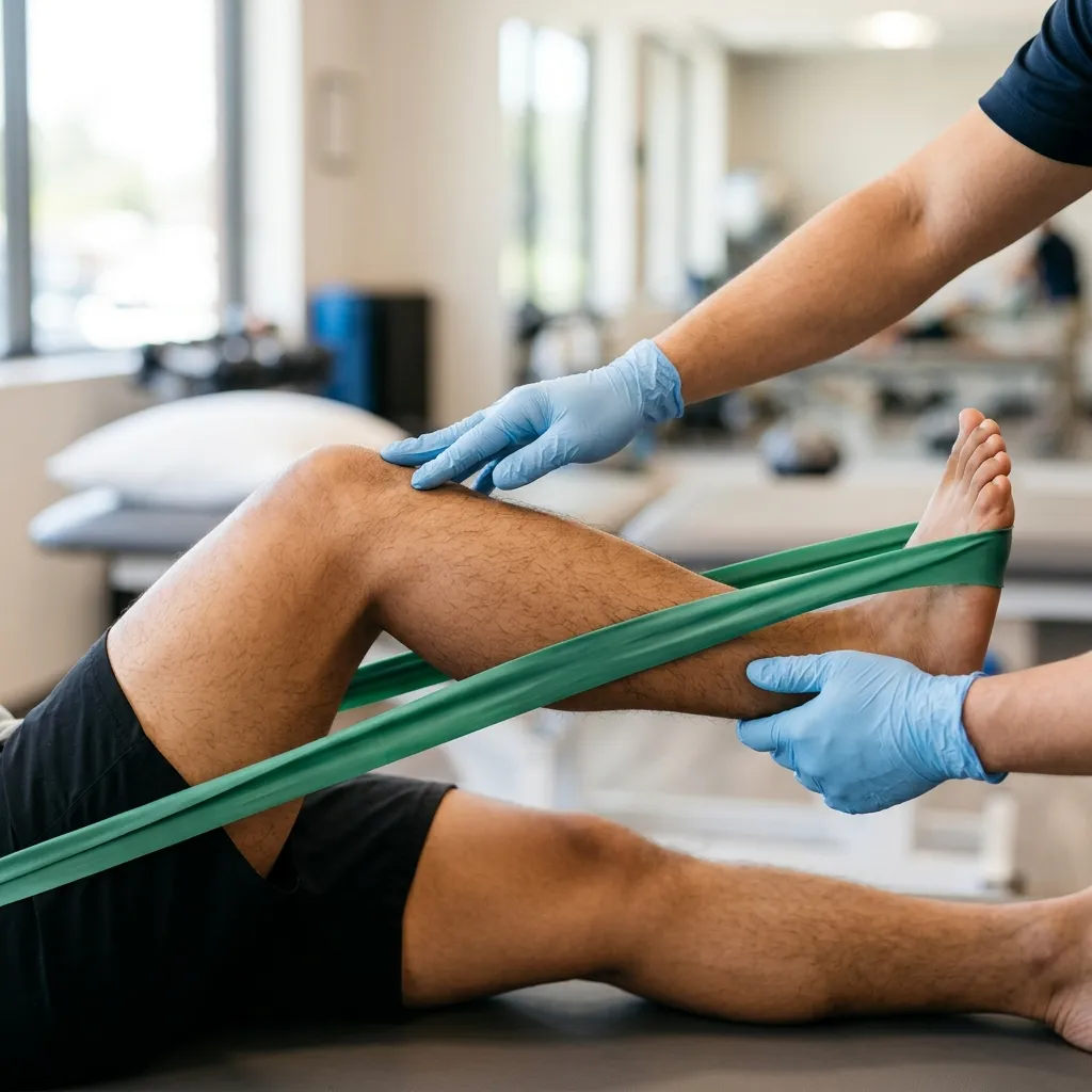
- — The surgeon builds the structural foundation; physical therapy rebuilds the muscular support and movement function.
- — Early, controlled movement prevents the formation of crippling scar tissue and restores joint mobility safely.
- — The timeline for returning to sport after ACL surgery relies on passing functional strength and control tests, not just counting weeks.
- — Therapeutic ultrasound and targeted exercises manage pain and wake up 'inhibited' muscles without risking the surgical repair.
Why Early Movement is Safe and Necessary
In my clinic, I explain that the surgeon builds the structure, but rehabilitation builds the function. The hardware or graft is actually strongest right after surgery. Moving early does not break the repair; it prevents crippling scar tissue and stiffness that can permanently limit your motion. The primary goal of post-operative rehabilitation is to respect tissue healing while safely reintroducing mechanical load.
When a joint undergoes surgery, the body initiates a profound inflammatory cascade. This swelling is a natural part of the healing process, but it introduces an immediate functional threat: Arthrogenic Muscle Inhibition. Because the joint is swollen and painful, the central nervous system reflexively shuts down the motor pathways to the surrounding muscles—particularly the quadriceps in the knee. Patients often describe their leg as feeling "heavy," "dead," or experiencing ngawit. This is not structural muscle damage; it is a neurological block. Our first clinical priority is to override this block through early, active muscle contraction to "wake up" the joint.
Current systematic reviews confirm that routine diagnostic imaging post-surgery or for non-traumatic joint pain is discouraged unless specific severe pathology is suspected, as it often leads to over-medicalization (Cuff et al., 2020; PMID: 32755058). Instead of relying on routine X-rays to check the hardware, we assess clinical progression: Is the swelling reducing? Is the range of motion improving? Is the muscle firing?
Contrarian Physician Myth 1: The "Complete Rest" Myth
A pervasive and dangerous myth among patients in the Philippines is that after joint surgery, you must completely immobilize the limb until it stops hurting. This "bed rest" approach is a biomechanical failure. Complete rest allows fibrinous scar tissue to organize and cement the joint capsule in place, leading to arthrofibrosis—a permanently frozen joint. Early, controlled movement pumps out edema, prevents the capsule from shrinking, and preserves your surgical investment.
Knee Replacement Rehab Protocol: Timeline and Expectations
I tell patients to expect a 6 to 12-week intensive recovery phase after a Total Knee Replacement (TKR). The absolute first priority is achieving full knee extension (straightening) within the first two weeks, followed by knee flexion (bending). Normal walking requires this extension; without it, you will have a permanent limp and compensatory back pain.
A knee replacement removes the arthritic surfaces and replaces them with metal and high-grade plastic components. However, the surrounding soft tissues—the ligaments, tendons, and muscles—must adapt to this new joint. When a patient lacks full extension (the ability to lock the knee perfectly straight), the quadriceps must work overtime simply to keep the person standing upright. This leads to profound fatigue and a shortened stride length.
Our protocol begins with immediate Quad Sets and Heel Slides. The patient tightening the thigh muscle to push the back of the knee flat against the bed forces the leg into terminal extension. The patient then actively slides the heel toward the buttocks to stretch the joint capsule into flexion. If pain is severely limiting this critical mobility window, evidence supports the use of therapeutic ultrasound to reduce local inflammation. Ultrasound therapy is significantly more effective than other interventions for reducing pain and aiding rehabilitation in knee conditions, serving as a strong adjunct to mobility training (Guan et al., 2024; PMID: 39170758).
Shoulder Rehab and Avoiding 'Frozen Shoulder'
After shoulder surgery, stiffness is your biggest enemy. I focus on early, controlled, passive motion to prevent the joint capsule from freezing. While the tendon or labrum heals, we must keep the joint gliding, balancing protection of the repair with the necessity of movement.
The shoulder is a highly mobile, complex joint that relies heavily on its muscular envelope (the rotator cuff) for stability. After a rotator cuff repair or shoulder replacement, the operated tissues are vulnerable to tensile stress. However, if the shoulder is kept immobilized in a sling for too long without controlled movement, the capsule thickens and contracts, resulting in an iatrogenic "Frozen Shoulder" (adhesive capsulitis).
To prevent this, we introduce Passive Range of Motion (PROM) exercises immediately. During PROM, the physical therapist or the patient's healthy arm does all the physical work to move the surgical arm. The muscles of the operated shoulder remain completely relaxed (no active contraction), which protects the surgical repair from tearing while safely stretching the capsule. As the tissue heals over several weeks, we progressively transition to Active-Assisted Range of Motion (AAROM), and eventually, Active strengthening.
Contrarian Physician Myth 2: The "No Pain, No Gain" Myth in Shoulder Rehab
While some discomfort is expected during joint rehabilitation, aggressive, painful forcing of a shoulder joint after surgery triggers a protective inflammatory response. If physical therapy is intensely painful, the patient’s muscles will involuntarily guard and spasm, creating more stiffness and potentially compromising the repair. Rehabilitation must respect the tissue's irritability level; we push to the edge of restriction, not through searing pain.
ACL Recovery Timeline: From Surgery to Sport
An ACL recovery timeline is not just about time; it is about strength and biomechanical control. I do not clear athletes based on weeks alone. You must pass functional tests—like single-leg hops and deceleration control—before returning to sports, which typically takes 9 to 12 months of progressive, staged rehabilitation.
The Anterior Cruciate Ligament (ACL) is a critical stabilizer of the knee, preventing the shin bone from sliding forward and providing rotational control. During an ACL reconstruction, the surgeon uses a graft (from your patellar tendon, hamstring, or a donor) to create a new ligament. The crucial physiological reality that patients misunderstand is the process of "ligamentization." Over the first 6 to 12 weeks, the new graft actually becomes weaker as its cells remodel and vascularize, before it slowly begins to strengthen.
Therefore, early rehab focuses strictly on resolving swelling, restoring full extension, and regaining basic quadriceps activation safely. Mid-stage rehab builds heavy strength and muscle hypertrophy. Late-stage rehab—often completely skipped by eager athletes—focuses on neuromuscular control: teaching the body how to land from a jump safely, decelerate rapidly, and cut laterally without the knee caving inward (dynamic valgus).
If patients attempt to shortcut this timeline and return to sports prematurely without passing rigorous biomechanical testing, the risk of a secondary ACL tear skyrockets.
Tactical Home Care: The Safe Alternative to Rest
The most common generic advice given to post-surgical patients in provincial settings is to "rest until the doctor sees you next month." This is a biomechanical fail.
Instead of complete immobilization, we institute a safe home care pivot: Immediate Isometrics and Active Pumping.
- Quad Sets: Tighten your thigh muscle to push the back of your knee flat into the bed. Hold for 5 seconds. This simple isometric contraction safely loads the quadriceps, preventing atrophy without moving the joint or stressing surgical repairs.
- Ankle Pumps: Vigorously point and flex your toes. This uses the calf muscles to pump venous blood and lymphatic fluid out of the leg, dramatically reducing swelling and lowering the risk of deep vein thrombosis (DVT).
- Heel Slides: Slowly slide your heel toward your buttocks to gently bend the knee, then straighten it. This prevents the capsule from shrinking.
These home exercises bridge the gap between clinic visits. In rare cases where extreme inflammation prevents any active loading, a targeted, ultrasound-guided corticosteroid injection might be considered to provide a short-term window of relief. Evidence suggests that while corticosteroid injections offer short-term joint pain relief, they must be used judiciously to avoid cartilage degradation; they buy a window of time for the patient to tolerate physical therapy (Benzon et al., 2025; PMID: 40015722).
Post-Surgical Care at TeraCare Clinic in Vigan
Post-surgical rehabilitation under Dr. Ben Paolo C. Rabara at TeraCare Clinic combines physiatric oversight, pain management, and certified physical therapy protocols. We understand the unique challenges faced by patients in Ilocos Sur, including the difficulty of traveling frequently for therapy.
Our approach focuses on empowering you. We assess your joint’s tissue state, monitor for surgical red flags (like infection or DVT), and design a progressive rehabilitation blueprint that integrates supervised clinical sessions with a highly structured home exercise program. We ensure that you are not just recovering, but actively rebuilding your strength to return to the activities you love.
Rebuild Your Joint Function Safely
Recovering from joint surgery requires precise, timed movement. A physiatric assessment helps identify your specific deficits, safely manages post-operative pain, and establishes a clear timeline for your physical therapy progression.
References & Clinical Evidence
- [1] Guan H, Wu Y, Wang X, Liu B, et al. Ultrasound therapy for pain reduction in musculoskeletal disorders: a systematic review and meta-analysis. Ther Adv Chronic Dis. 2024;15:20406223241267217. PMID: 39170758.
- [2] Cuff A, Parton S, Tyer R, Dikomitis L, et al. Guidelines for the use of diagnostic imaging in musculoskeletal pain conditions affecting the lower back, knee and shoulder: A scoping review. Musculoskeletal Care. 2020;18(4):546-554. PMID: 32755058.
- [3] Benzon HT, Provenzano DA, Nagpal A, Souza D, et al. Use and safety of corticosteroid injections in joints and musculoskeletal soft tissue: guidelines from the ASRA, AAPM, ASIPP, and IPSIS. Reg Anesth Pain Med. 2025;50(2):83-102. PMID: 40015722.
* Clinical references are provided to support the medical claims made in this article. TeraCare adheres to evidence-based practices in physical medicine and rehabilitation.

Dr. Ben Rabara
Dr. Ben Rabara is a Board-Certified Physiatrist specializing in Physical Medicine and Rehabilitation. He focuses on non-surgical, precision treatments for musculoskeletal conditions, utilizing advanced diagnostics like MSK Ultrasound.
Medical Disclaimer: The information provided in this article is for educational purposes only and does not substitute for professional medical advice, diagnosis, or treatment. Always consult a qualified physician for your specific health conditions.