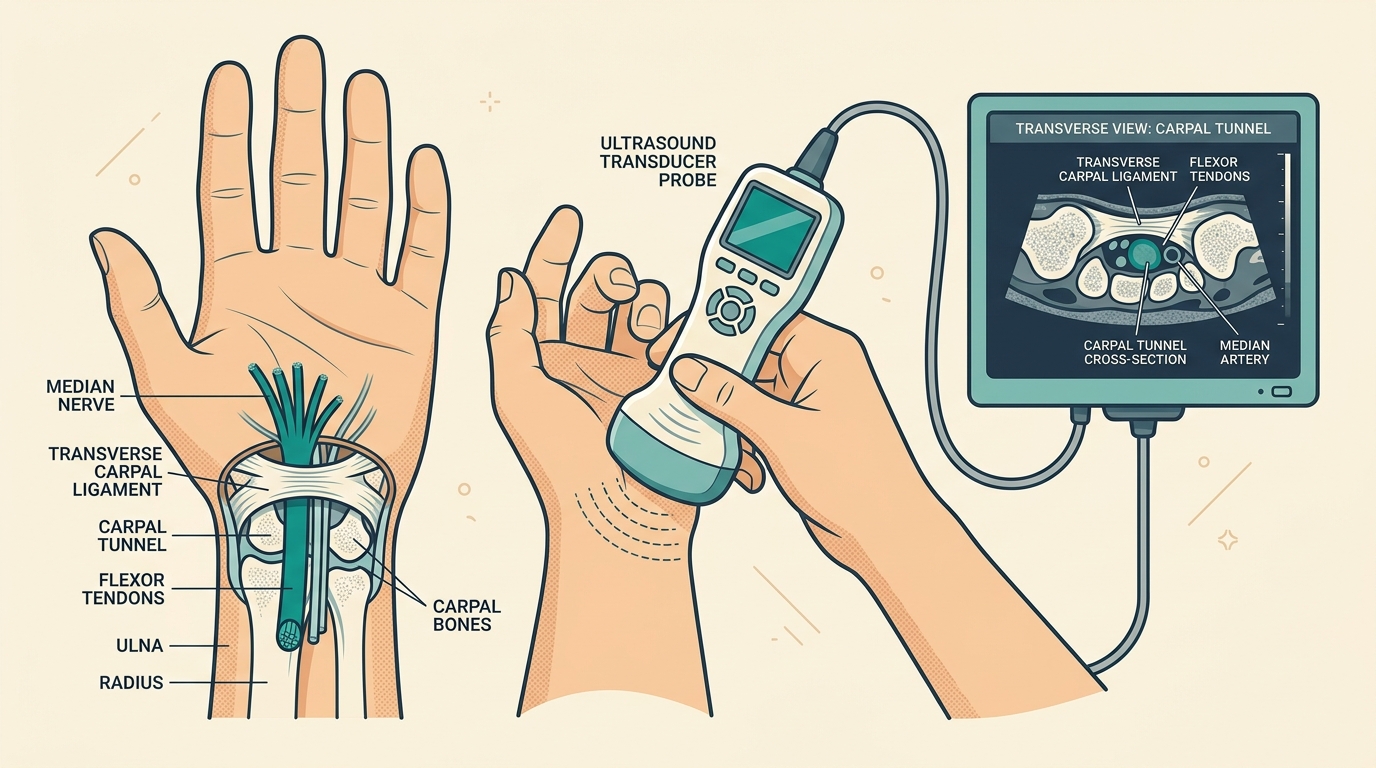
- — Waking up at 3:00 AM with numb, tingling hands that require 'shaking out' is the classic hallmark of Carpal Tunnel Syndrome (CTS).
- — Sleeping with flexed wrists increases pressure inside the carpal tunnel from a normal baseline of 2.5 mmHg to over 30 mmHg, collapsing microvascular blood flow.
- — Point-of-Care MSK Ultrasound (POCUS) provides instant bedside diagnosis by measuring median nerve cross-sectional area (cutoff 10 mm2), avoiding painful EMG nerve shocks.
- — Clinical provocation tests like Phalen wrist flexion for 60 seconds and Durkan compression for 30 seconds confirm nerve entrapment beneath the 2.5 mm thick ligament.
Nighttime hand numbness is rarely a subtle discomfort. It is defined as a nocturnal sensory disturbance characterized by severe tingling, numbness, and burning pain that violently wakes individuals from deep sleep. Waking up at 3:00 AM with hands that feel completely dead, impossibly stiff, or filled with intense, throbbing "pins and needles" is an alarming experience. Many patients describe desperately shaking their wrists, hanging their arms off the side of the bed, or running warm water over their fingers just to bring sensation back.
While many people attribute this terrifying numbness to "poor blood circulation" or simply sleeping awkwardly on an arm, the true underlying cause is almost always median nerve compression inside the carpal tunnel (Carpal Tunnel Syndrome). The psychological toll of this condition is often overlooked. Night after night of interrupted sleep leads to chronic exhaustion. During the day, patients experience the devastating frustration of losing their grip strength—dropping coffee cups, struggling to turn doorknobs, or failing to button their shirts.
In my physical medicine and rehabilitation clinic, I evaluate patients presenting with nocturnal hand numbness every week. When a patient arrives describing how they wake up with a stiff, heavy, and weak hand accompanied by a sharp pain, dull ache, radiating discomfort, or burning sensation, I immediately look for objective, structural signs of median nerve entrapment. Below, we examine the precise anatomy of the carpal tunnel, explain the biomechanics of why sleep triggers such severe pressure spikes, detail how to physically test your own wrist at home, and outline why high-resolution ultrasound is the new gold standard for diagnosis.
Anatomy & Biomechanics: The Structural Bottleneck
To understand why your hand goes numb, you must first understand the structural engineering of your wrist. The carpal tunnel is a rigid, unyielding passageway located on the palm side of your wrist.
Think of it as a subterranean concrete tunnel. Its floor and walls are formed by the hard carpal wrist bones. Its roof is sealed shut by a thick, incredibly strong band of connective tissue called the transverse carpal ligament (flexor retinaculum). Passing through this extremely narrow, crowded tunnel are ten vital structures: nine flexor tendons (which pull your fingers into a fist) and one single, highly sensitive electrical cable: the median nerve.
Because the walls of the tunnel are made of bone and dense ligament, the carpal tunnel is a "closed compartment." It cannot stretch or expand. If the tendons swell from repetitive typing, heavy lifting, or hormonal changes, the pressure inside the tunnel skyrockets. The tendons are tough and can withstand the pressure, but the soft, fragile median nerve gets crushed against the ligamentous roof.
The Physiology of Sleep: Ischemic Capillary Collapse
So why does this compression become so overwhelmingly painful specifically while you are sleeping? The answer lies in your sleeping posture and the microscopic blood supply of the nerve.
During the day, when your wrist is straight (in a neutral position), the hydrostatic pressure inside a healthy carpal tunnel is very low—typically between 2.5 to 5 mmHg. However, during deep sleep, the vast majority of people unconsciously curl their wrists inward into a flexed fetal position.
- The Pressure Spike: Bending the wrist to 90 degrees acts like folding a garden hose. It dramatically reduces the volume of the carpal tunnel, instantly spiking the internal pressure from a normal 3 mmHg to a dangerous 30 to 50 mmHg.
- Ischemic Capillary Collapse: Nerves have their own microscopic blood vessels called the vasa nervorum. When the pressure inside the tunnel exceeds 30 mmHg, these tiny blood vessels are physically squeezed shut. Blood stops flowing. The nerve is instantly starved of oxygen—a state called ischemia.
- Axonal Transport Failure: Deprived of oxygen, the internal transport system of the nerve (axoplasmic flow) grinds to a halt. The nerve panics and begins misfiring, sending erratic, high-frequency ectopic signals to the brain. Your brain interprets this neurological distress as intense burning, tingling, and deep aching pain.
The Pathognomonic "Flick Sign"
The classic human response to this nocturnal ischemia is the Flick Sign. When the pain wakes you up, your immediate, unconscious reaction is to violently shake or "flick" your hands, much like you are trying to shake water off your fingers.
Why does this work? Shaking the hand forces the wrist out of its curled position, dropping the internal pressure back down. It also utilizes centrifugal force to artificially drive venous blood back into the starved capillaries. Clinical studies demonstrate that the presence of the Flick Sign has a diagnostic sensitivity exceeding 93% for Carpal Tunnel Syndrome. If you are shaking your hands awake at 3:00 AM, you almost certainly have CTS.
Differential Diagnosis: Is It Really Carpal Tunnel?
Not all hand numbness originates at the wrist. It is absolutely vital to differentiate Carpal Tunnel Syndrome from other nerve entrapment conditions to avoid incorrect treatments or unnecessary surgeries. The key to the puzzle is the exact anatomical map of your numbness.
The median nerve provides sensation to a very specific territory: the thumb, the index finger, the middle finger, and exactly one-half (the radial side) of the ring finger.
The Pinky Finger Rule: The median nerve does not wire the pinky finger. If your pinky finger (little finger) is completely normal, but your thumb and middle fingers are numb, you have Carpal Tunnel Syndrome. However, if your pinky finger and the outside of your ring finger are numb, the entrapment is happening at the ulnar nerve—usually at the elbow (Cubital Tunnel Syndrome).
| Condition | Primary Nerve Involved | Symptom Distribution Pattern | Key Distinguishing Hallmark |
|---|---|---|---|
| Carpal Tunnel Syndrome | Median Nerve (Wrist) | Thumb, Index, Middle, & Half of Ring Finger | Nocturnal waking; Pinky finger completely spared |
| Cervical Radiculopathy | C6 / C7 Nerve Roots (Neck) | Radiating arm pain down into specific fingers | Looking up or rotating the neck triggers an electrical shock down the arm |
| Cubital Tunnel Syndrome | Ulnar Nerve (Elbow) | Little Finger (Pinky) & Ulnar Ring Finger | Numbness triggered by sleeping with elbows bent tight |
| Peripheral Neuropathy | Distal Microvascular Nerves | Symmetrical "Glove & Stocking" distribution | Affects both hands and both feet equally; highly correlated with Diabetes |
Clinical Provocation Tests: Physical Exams that Prove the Pinch
Before relying on expensive imaging, we perform targeted physical maneuvers in the clinic designed to artificially recreate the pressure spike and provoke median nerve symptoms. You can gently test these at home to gauge your likelihood of entrapment:
- Phalen's Wrist Flexion Test: Hold your wrists in complete, unforced flexion by pressing the backs of your hands together (fingers pointing down) for a strict **60 seconds**. Pushing the median nerve against the transverse carpal ligament reproduces the nocturnal numbness or tingling in the thumb and index finger. A positive result within 60 seconds strongly indicates compression.
- Durkan's Carpal Compression Test: The physician applies direct, forceful manual thumb pressure exactly over the carpal tunnel for 30 seconds. This is recognized as the most sensitive and specific physical examination test for early CTS, as it directly mimics the high-pressure ischemic state.
- Tinel's Percussion Sign: Lightly tapping (percussing) over the volar surface of the wrist directly above the carpal tunnel. A positive test produces an electric-shock sensation that rapidly shoots into the median-innervated fingers.
The Gold Standard: POCUS Ultrasound Diagnostics
The definitive physiological diagnosis of Carpal Tunnel Syndrome can be verified through an Electromyography and Nerve Conduction Velocity (EMG/NCV) test to calculate the microsecond slowdown of the nerve signal.
At our clinic, we pair electrodiagnostics with high-resolution Point-of-Care MSK Ultrasound (POCUS). By placing the ultrasound probe directly on the wrist, we can visually inspect the structural integrity and cross-sectional area of the nerve in real-time.
Key sonographic diagnostic parameters include:
- Median Nerve Cross-Sectional Area (CSA): This is the most critical metric. We measure the circumference of the nerve at the inlet of the carpal tunnel (near the pisiform bone). A healthy median nerve measures around 6 to 9 mm². If the nerve is trapped, it swells up like a dammed river just before the pinch. A cross-sectional area measuring 10 mm² or larger confirms significant nerve swelling and structural entrapment with over 85% sensitivity.
- The "Notch Sign" (Hourglass Deformity): By turning the ultrasound probe lengthwise, we can see the nerve getting physically crushed flat as it dives under the transverse carpal ligament, creating an hourglass shape.
- Palmar Bowing of Transverse Carpal Ligament: The dangerously high internal tunnel pressure physically pushes the thick ligament roof outward toward the palm. Palmar displacement exceeding 2.5 mm indicates severe capsular tightness.
The Path to Non-Surgical Relief
If you are waking up with numb hands, ignoring the problem is incredibly dangerous. Prolonged ischemic pressure (>50 mmHg) eventually leads to permanent nerve death, irreversible loss of protective sensation, and atrophy (wasting away) of the thumb muscles.
The very first conservative step is to purchase a rigid night splint. This splint holds your wrist in a neutral position (0° to 15° of extension) while you sleep, preventing the unconscious curling that spikes the tunnel pressure. For mild cases, wearing a night splint can resolve the nocturnal ischemia entirely.
However, if splinting fails and the ultrasound confirms significant nerve swelling (CSA > 10 mm²), intervention is required to prevent permanent damage. Before resorting to invasive open release surgery—which involves slicing the transverse carpal ligament in half with a scalpel—we offer state-of-the-art non-surgical options.
At TeraCare Physical Medicine & Rehabilitation Clinic in Vigan City, Dr. Ben Rabara utilizes Ultrasound-Guided Nerve Hydrodissection. Using a "liquid scalpel," we inject a precise stream of therapeutic fluid (D5W) perfectly around the median nerve. The fluid pressure safely peels away the constricting scar tissue, mechanically expanding the carpal tunnel space and instantly un-pinching the nerve without a single surgical cut.
For deeper details on how real-time fluid injection releases trapped nerves without surgery, read our Nerve Hydrodissection Science Guide. For a full comparison between open surgery vs. hydrodissection, consult our Alternatives to Carpal Tunnel Surgery Guide.
Schedule Your Nerve Evaluation in Vigan City
Stop waking up with numb hands. Book an expert ultrasound nerve scan and evaluation with Dr. Ben Rabara today.
Clinical Realities of Median Nerve Entrapment
Understanding nocturnal pressure surges, ischemic nerve conduction, and early diagnosis.
Nocturnal Pressure Surge
Flexing wrists during sleep spikes carpal tunnel pressure past 30 mmHg, shutting down venous micro-circulation to median nerve fibers.
Anatomical Sensory Distribution
Numbness selectively strikes the thumb, index, middle, and ring fingers while completely sparing the pinky finger.
Bedside POCUS Diagnostic Standard
Ultrasound measures nerve swelling (cross-sectional area >= 10 mm²) instantly without needing uncomfortable needle EMG electrical shocks.
References & Clinical Evidence
- [1] Elsawy AM, et al. Diagnostic accuracy of point-of-care ultrasound versus clinical physical examination for detection of nerve compression. BMC Musculoskelet Disord. 2023. PMID: 10257372.
- [2] Dainese P, Wyngaert KV, De Mits S, Wittoek R, Van Ginckel A, Calders P. Association between nerve compression and neuropathic pain: a systematic review. Osteoarthritis Cartilage. 2022 Apr;30(4):516-534. doi: 10.1016/j.joca.2021.12.003. PMID: 34968719.
- [3] Sukerkar PA, Doyle Z. High-Resolution Musculoskeletal Ultrasound of Peripheral Nerves. Radiol Clin North Am. 2022 Jul;60(4):583-596. doi: 10.1016/j.rcl.2022.03.004. PMID: 35672093.
- [4] Aiyer R, et al. Therapeutic Ultrasound and Hydrodissection for Chronic Pain Management: A Systematic Review. Pain Med. 2020. PMID: 31095336.
* Clinical references are provided to support the medical claims made in this article. TeraCare adheres to evidence-based practices in physical medicine and rehabilitation.

Dr. Ben Rabara
Dr. Ben Rabara is a Board-Certified Physiatrist specializing in Physical Medicine and Rehabilitation. He focuses on non-surgical, precision treatments for musculoskeletal conditions, utilizing advanced diagnostics like MSK Ultrasound.
Medical Disclaimer: The information provided in this article is for educational purposes only and does not substitute for professional medical advice, diagnosis, or treatment. Always consult a qualified physician for your specific health conditions.