- — Temporary numbness from sleeping awkwardly resolves in minutes; chronic hand numbness waking you at night indicates true nerve entrapment.
- — Carpal Tunnel Syndrome involves mechanical compression of the median nerve at the wrist, selectively sparing the pinky finger.
- — An EMG-NCV test acts as an electrical speedometer, pinpointing the exact location, severity, and physiological impact of nerve damage.
- — Ulnar nerve entrapment (Cubital Tunnel) chokes a different nerve pathway, causing numbness isolated to the pinky and outer ring fingers.
- — Delaying diagnostics allows nerve injury to progress from sensory tingling to irreversible motor axon death and muscle wasting (thenar atrophy).
It starts as a minor annoyance: a mild tingling in your index and middle fingers while driving, or a slight stiffness when buttoning your shirt. But soon, it escalates. You begin waking up in the middle of the night with your entire hand feeling completely numb, cold, and burning. You vigorously shake your hand over the edge of the bed, waiting for the "electricity" to return, wondering if you simply slept on your arm awkwardly.
In my clinical practice as a Physical Medicine & Rehabilitation (PM&R) physician, I meet patients every single week who have spent months—sometimes years—attributing this progressive hand numbness to "sleeping wrong" or poor blood circulation. Many have consumed boxes of Vitamin B-complex or worn generic wrist braces bought online, only to watch their grip strength slowly deteriorate.
The truth is, nerves are the electrical wiring of your body. When a wire is pinched, it doesn't just cause temporary pain; it undergoes progressive, structural degradation. To protect your hand function, you must stop guessing. Understanding whether your hand numbness is a transient sleeping posture or a progressive entrapment neuropathy requires the clinical clarity of an Electromyography and Nerve Conduction Velocity (EMG-NCV) study.
Is It Just a 'Sleeping Arm' or True Nerve Entrapment?
Numbness in hands often signals progressive median nerve compression rather than a simple temporary awkward sleeping position. When fingers regularly wake you up with a burning, "asleep" sensation, the transverse carpal ligament may be mechanically choking your nerve. Delaying a clinical diagnostic assessment allows subclinical axonal damage to quietly progress toward irreversible hand weakness.
To understand why your hand falls asleep, we must separate two entirely different physiological events: transient neuropraxia and chronic entrapment neuropathy.
When you sleep awkwardly on your arm, you compress the blood vessels supplying a nerve, or squeeze the nerve pathway directly. This blocks the nerve’s ability to transmit electrical impulses. Once you wake up, change positions, and shake your arm, blood flow restores, the nerve "wakes up" with a brief pins-and-needles sensation, and normal function returns within seconds or minutes. This is a temporary, benign physiological event known as transient neuropraxia.
In contrast, chronic nerve entrapment is a structural bottleneck. In conditions like Carpal Tunnel Syndrome, the median nerve is physically squeezed 24 hours a day inside a narrow, rigid anatomical canal in your wrist. Even when you are awake, typing, or resting, the nerve is under constant mechanical stress. Nighttime merely exacerbates this pressure because we naturally bend our wrists while sleeping, skyrocketing internal canal pressure and choking the nerve's blood supply.
If you experience any of the following patterns, your numbness is likely not just a temporary sleeping pinch:
- Symptom Consistency: The numbness occurs almost every single night, or during routine daytime activities like holding a phone, driving a motorcycle, or gripping a coffee cup.
- Clumsiness: You find yourself dropping lightweight objects, struggling to open jar lids, or experiencing difficulty buttoning your clothes.
- Selective Numbness: The numbness selectively affects your thumb, index, middle, and half of your ring finger, while your pinky finger feels completely normal.
What is Carpal Tunnel Syndrome? The Wrist Bottleneck
Carpal tunnel syndrome symptoms arise when swelling or structural narrowing inside the rigid wrist canal squeezes the median nerve. This nerve controls sensation in the thumb, index, middle, and half of the ring finger, as well as the thumb's base muscles. Without objective electrical speed mapping, physicians cannot distinguish wrist-level compression from higher bottlenecks in the elbow or neck.
To visualize Carpal Tunnel Syndrome (CTS), think of your wrist as a busy, rigid highway tunnel. The floor and walls of this tunnel are formed by the hard carpal bones of your wrist, while the ceiling is sealed by a tough, inelastic band of tissue called the transverse carpal ligament (or flexor retinaculum).
Squeezed inside this tight space are ten critical structures: nine flexor tendons that allow you to bend your fingers, and one single, highly sensitive nerve—the median nerve.
Because the walls and ceiling of the carpal tunnel are completely rigid, they cannot expand. If the sheaths surrounding your flexor tendons become swollen or inflamed—due to repetitive hand movements, chronic typing, driving, metabolic shifts (like diabetes, thyroid disease, or pregnancy), or localized arthritis—the volume inside the tunnel increases. Since the tunnel cannot expand, the extra pressure is forced directly onto the softest, most vulnerable structure: the median nerve.
"The median nerve is like a soft garden hose. When the tendons swell, they step on the hose. The water—which represents your nerve's sensory and motor signals—slows down or stops flowing entirely."
In the early stages of this squeeze, the nerve's outer protective coating (the myelin sheath) is compressed. This triggers the classic symptoms of tingling, burning, and pain. If the squeeze remains untreated, the pressure chokes off the microscopic blood vessels supplying the nerve fibers (axons) themselves, leading to focal nerve ischemia, permanent structural damage, and eventual muscle wasting.
Why Do You Need an EMG-NCV Test for Hand Numbness?
An electromyography and nerve conduction velocity study serves as a precision speedometer for your hand's electrical highway. The NCV portion calculates electrical conduction velocity and latency across the wrist to pinpoint the exact site of signal delay. The needle EMG portion evaluates whether nerve fibers are actively dying, revealing if muscle tissue is losing its neural connections.
When you consult a doctor for hand numbness, they may perform simple physical maneuvers in the clinic, such as tapping on your wrist (Tinel's Sign) or holding your wrists in a bent position (Phalen's Test) to see if they can trigger your symptoms. While these tests are helpful screening tools, they are highly subjective and frequently yield false positives or false negatives.
Furthermore, standard imaging studies like X-rays or MRIs are structurally focused. An X-ray can show your wrist bones, and an MRI can take a beautiful picture of your soft tissues, but neither can tell you how well the nerves are actually functioning. They cannot prove if electricity is flowing smoothly through the wires.
This is why an EMG-NCV test is the absolute clinical gold standard. It provides objective, mathematical proof of your nerve health by performing two distinct diagnostic functions:
- Nerve Conduction Velocity (NCV): By placing surface electrodes on your skin and delivering a tiny, completely safe electrical stimulus, we measure exactly how fast (in meters per second) and how strong (in microvolts) your nerve signals travel. If the signal slows down significantly as it crosses the wrist, we have objective proof of a bottleneck.
- Electromyography (EMG): By inserting a microscopic, sterile needle electrode into specific muscles controlled by the median nerve, we "listen" to the internal electrical activity of your muscle fibers. If the nerve is severely pinched and failing to supply the muscle, the muscle will emit abnormal, chaotic electrical waves at rest (such as fibrillation potentials and positive sharp waves). This tells us that nerve fibers are actively dying.
With this data, Dr. Rabara can classify your carpal tunnel syndrome as Mild, Moderate, or Severe. This classification is critical because it dictates your entire treatment plan: mild cases recover quickly with simple nighttime splinting, moderate cases benefit immensely from precision nerve hydrodissection, while severe cases showing muscle denervation may require a surgical release to prevent permanent loss of hand function.
Carpal Tunnel vs. Ulnar Nerve Entrapment: Differentiating the Bottlenecks
Hand numbness manifests differently depending on which nerve path is compressed, distinguishing median nerve carpal tunnel from ulnar nerve cubital tunnel entrapment. Carpal tunnel selectively spares the pinky finger, whereas ulnar nerve compression at the elbow isolates numbness to the pinky and outer ring finger. Electrodiagnostic mapping mathematically separates these distinct nerve channels to guide targeted rehabilitation or surgical relief.
Your hand is supplied by more than one nerve pathway. While the median nerve travels through the carpal tunnel, another major pathway—the ulnar nerve—runs along the inner side of your elbow (the "funny bone" area) and down into the outer side of your wrist through a small channel called Guyon's Canal.
When the ulnar nerve is compressed—most commonly at the elbow, a condition known as Cubital Tunnel Syndrome—it also causes hand numbness. However, the distribution of symptoms is completely different.
If you are experiencing ulnar nerve entrapment, your numbness and tingling will be localized to your pinky finger and the outer half of your ring finger. You will often feel a deep ache on the inner side of your elbow, and your hand may feel weak when you try to spread your fingers apart or grip objects tightly.
Without an EMG-NCV test, it is very common to misdiagnose ulnar nerve entrapment as carpal tunnel syndrome, or vice versa. In worse cases, a patient might have a "Double Crush" syndrome, where nerves are compressed at multiple levels simultaneously—such as a pinched nerve in the neck (Cervical Radiculopathy) co-existing with carpal tunnel syndrome at the wrist.
Treating only the wrist when the root cause is in the neck or elbow is a recipe for treatment failure. An EMG-NCV study maps the entire length of the nerves, systematically testing the neck, shoulder, elbow, and wrist to trace the exact source of your pain.
Self-Care vs. Professional Diagnostic Assessment
To help you visualize the critical differences between ignoring hand numbness (or relying on unverified home remedies) and undergoing a professional electrodiagnostic evaluation, consider this clinical comparison:
| Clinical Metric | Self-Care & Wait-and-See | Professional EMG-NCV Diagnostic |
|---|---|---|
| Diagnostic Accuracy | Subjective guessing based on generic online articles or symptoms. | 100% Objective: Maps exact speed, location, and severity of nerve pinch. |
| Risk of Muscle Wasting | High: Undetected severe compression quietly kills motor axons distally. | Prevented: Identifies early denervation before physical wasting begins. |
| Treatment Direction | Blind application of random braces, supplements, and massage. | Targeted: Guides exact sequencing (splints vs hydrodissection vs surgery). |
| Long-Term Outcomes | Poor: Often leads to chronic pain, permanent weakness, or delayed surgery. | Excellent: High recovery rate via customized, non-surgical rehabilitation plans. |
Preventing Permanent Nerve Damage: The Loss of Grip Strength
Chronic nerve compression eventually transitions from transient sensory numbness to irreversible motor axon death and muscle wasting. The thenar eminence—the muscular pad at the base of the thumb—flatlines when the median nerve is strangled too long. Getting a timely EMG-NCV test allows patients to intervene with conservative therapies before losing fine motor coordination and grip strength.
Nerves are composed of thousands of individual microscopic fibers bundled together, much like a thick telecommunications cable. These fibers are divided into two main categories: sensory fibers (which transmit sensations like touch, temperature, and pain to your brain) and motor fibers (which carry signals from your brain to your muscles, commanding them to contract).
Sensory fibers are thinner and have less protective wrapping, making them highly sensitive to pressure. This is why the early stages of carpal tunnel syndrome are dominated by sensory symptoms: tingling, burning, and numbness. During this phase, the nerve is injured but still intact, and the damage is entirely reversible.
However, if the compression continues unmitigated, the pressure begins to choke the thicker, more resilient motor fibers. When these motor fibers are damaged, they can no longer transmit signals to the small muscles in your hand.
The most severe consequence of untreated carpal tunnel syndrome is thenar atrophy. The thenar eminence is the thick, fleshy muscle pad at the base of your thumb. It is what allows you to oppose your thumb to your other fingers, enabling you to pinch, write, hold a pen, grip a glass, or button your shirt.
When the motor axons of the median nerve die, the thenar muscles lose their electrical connection. Deprived of stimulation, the muscle fibers begin to shrink and waste away. Visually, the fleshy pad at the base of your thumb becomes completely flat or hollowed out.
Once thenar atrophy occurs, the damage is very difficult to reverse. Even if you undergo surgery later to release the ligament, the wasted muscle may never fully recover its volume or strength. This is why waiting until you lose grip strength or notice hand flatlining is a clinical tragedy—you must get an EMG-NCV test while the nerve damage is still in the reversible, sensory stage.
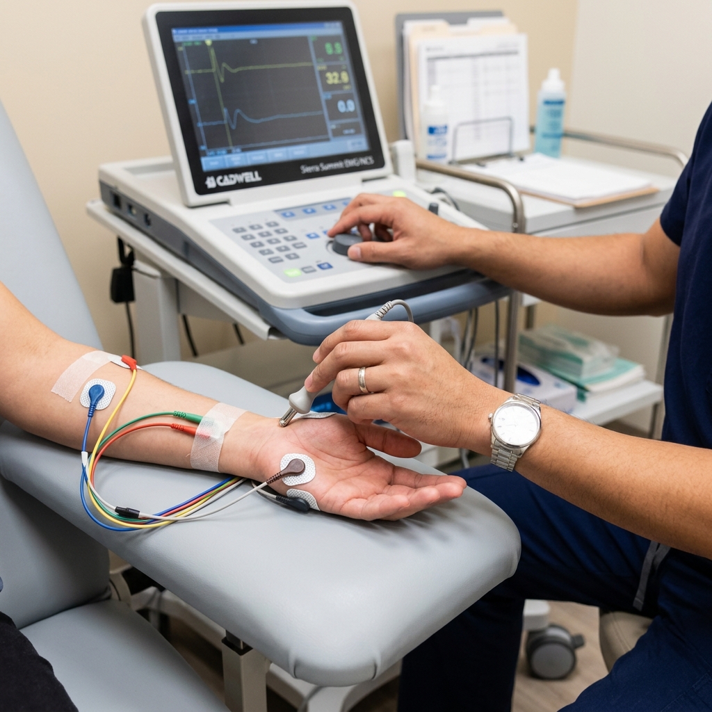
What Happens During an EMG-NCV Test? Overcoming the Fear
Electrodiagnostic testing consists of two sequential phases designed to analyze different aspects of neuromuscular health. The nerve conduction velocity (NCV) test applies small, static-like electrical pulses to the skin to measure signal speed. The electromyography (EMG) portion inserts a microscopic, sterile needle into specific hand and arm muscles to record silent or abnormal muscle waveforms.
It is entirely normal to feel anxious when you hear the words "electrical shocks" or "needles." Much of this anxiety comes from the unknown. Let us walk through exactly what happens during the procedure at TeraCare Vigan to demystify the experience:
Phase 1: The Nerve Conduction Study (NCV)
You will sit or lie down comfortably. Dr. Rabara will place small recording stickers (electrodes) on your fingers and palm. Using a handheld stimulator, he will deliver brief, mild electrical pulses to various points along your arm.
Each pulse lasts only a fraction of a millisecond. It feels like a sudden tapping sensation or a mild static electricity shock (similar to touching a metal doorknob after walking on a carpet). Your fingers may twitch involuntarily during the stimulation; this is a normal, healthy physical reflex. The machine records exactly how long the signal takes to travel from the stimulation point to the recording electrode, calculating your nerve speed to the microsecond.
Phase 2: The Needle Electromyography (EMG)
During this portion, Dr. Rabara will insert a microscopic, sterile needle electrode into specific muscles of your hand, forearm, and sometimes your shoulder.
Unlike a vaccination or blood draw needle, this diagnostic needle is incredibly thin, solid (it does not inject any chemicals or draw any blood), and acts purely as a microphone to listen to your muscle's natural electrical activity. You will feel a small pinch as it enters the muscle, similar to an acupuncture needle.
You will hear a static popping sound over the machine's speaker, which represents the sound of your muscle fibers contracting. Dr. Rabara will ask you to relax the muscle completely, and then contract it gently. He will observe the resulting electrical waveforms on a monitor, analyzing their amplitude, duration, and frequency.
The entire test takes between 30 to 45 minutes. There are absolutely no side effects, no sedation is required, and there is zero downtime. You can drive yourself home immediately, return to work, and resume your normal activities. The minor, localized muscle soreness you may experience resolves completely within a day.
Why Choose a Physiatrist for Your Nerve Test in Vigan City?
Physical Medicine and Rehabilitation specialists, or Physiatrists, are uniquely qualified to perform and interpret electrodiagnostic tests because they integrate diagnostics directly with functional recovery. Dr. Ben Rabara provides localized, precision EMG-NCV mapping in Vigan City, eliminating the need to travel to distant metropolitan centers. This local expertise ensures patients receive immediate, board-certified physical medicine guidance under one roof.
In many healthcare settings, electrodiagnostic studies are performed by a technician who simply prints out a raw report for another doctor to read weeks later. This fragmented approach often leads to diagnostic errors or missed clinical details.
At TeraCare Vigan, your entire EMG-NCV test is personally conducted, analyzed, and interpreted in real-time by Dr. Ben Paolo C. Rabara, a Board-Certified Physiatrist.
As a PM&R specialist, Dr. Rabara is trained to view the human body as an integrated functional system. He doesn't just look at the numbers on the screen; he correlates your electrical waveforms directly with your physical examination, your daily activities, your unique anatomy, and your functional goals.
Because he performs the test himself, he can adjust the diagnostic protocol in real-time. If he detects an unexpected signal at the wrist, he can immediately test the elbow or neck to rule out other compression points in a single session.
Furthermore, having an advanced diagnostic lab right here in Vigan City provides immense practical benefits for families in Ilocos Sur and Northern Luzon:
- No Travel Fatigue: You do not need to endure a exhausting, painful 7-hour road trip to Manila just to get your nerves mapped. You can get world-class, university-level diagnostic accuracy right in your home region.
- Seamless Transition to Treatment: Because your diagnostic doctor is also your treatment doctor, there is no waiting. The moment your test is complete, Dr. Rabara will sit down with you, explain your "Nerve Map," and immediately orchestrate a personalized recovery plan.
- Affordable & Accessible: We believe in absolute pricing transparency. Our diagnostic fees are highly competitive, we are fully accredited, and we provide complete documentation to support PhilHealth claims for diagnostic laboratory procedures.
Conclusion: Restore Your Hand Health Before the Decline
Do not wait for your hand to lose its strength, and do not ignore the nightly numbness that disrupts your sleep. Hand numbness is not an inevitable consequence of aging or hard work—it is a clear warning signal from your nervous system that a pathway is being starved of oxygen and blood flow.
A precision EMG-NCV diagnostic test at TeraCare Vigan takes less than an hour, but it provides the definitive answers you need to stop the progressive decline. Whether your symptoms require simple custom bracing, advanced nerve hydrodissection, or surgical referral, a proper diagnostic map is the essential first step toward reclaiming pain-free hand function.
Map Your Nerve Health Today
Stop guessing why your hand is falling asleep. Schedule a precision, board-certified EMG-NCV diagnostic study with Dr. Ben Rabara in Vigan City.
Clinical Review Note: This educational guide was drafted under direct physician supervision and thoroughly reviewed for electrodiagnostic accuracy, anatomical precision, and patient safety before publication.
References & Clinical Evidence
- [1] American Association of Neuromuscular & Electrodiagnostic Medicine (AANEM). (2020). Evidence-based guideline: Clinical utility of electromyography and nerve conduction studies in carpal tunnel syndrome.
- [2] Kasius, K. M., et al. (2022). Association of electrodiagnostic severity with clinical presentation and outcomes in carpal tunnel syndrome. Muscle & Nerve Journal.
- [3] Werner, R. A., & Andary, M. (2011/Re-evaluated 2021). Carpal tunnel syndrome: Pathophysiology and electrodiagnosis. Physical Medicine & Rehabilitation Clinics of North America.
- [4] Graham, B., et al. (2019/Current Standard). The American Academy of Orthopaedic Surgeons Systematic Review and Clinical Practice Guideline on the Management of Carpal Tunnel Syndrome.
* Clinical references are provided to support the medical claims made in this article. TeraCare adheres to evidence-based practices in physical medicine and rehabilitation.

Dr. Ben Rabara
Dr. Ben Rabara is a Board-Certified Physiatrist specializing in Physical Medicine and Rehabilitation. He focuses on non-surgical, precision treatments for musculoskeletal conditions, utilizing advanced diagnostics like MSK Ultrasound.
Medical Disclaimer: The information provided in this article is for educational purposes only and does not substitute for professional medical advice, diagnosis, or treatment. Always consult a qualified physician for your specific health conditions.