- — An MRI provides a structural snapshot (like a blueprint photo), whereas an EMG measures live functional electricity (like an electrician's multimeter).
- — Over 30% of completely healthy, pain-free individuals have herniated discs on their MRIs, which can lead to treating the wrong disc without an EMG.
- — Distal nerve testing—testing the legs or arms—is the only way to prove exactly which spinal nerve root (e.g., L4, L5, S1, or C6, C7) is structurally choked.
- — Drop foot is a severe motor symptom typically caused by L5 nerve root compression, demanding immediate EMG testing to gauge nerve survival.
- — An EMG-NCV test is essential before undergoing any spinal decompression or fusion to confirm the exact pain generator and avoid 'Failed Back Surgery Syndrome'.
It is a pain unlike any other: a sudden, hot, electric shock that shoots from your lower back, rips through your buttock, and travels all the way down the back of your leg into your foot. Every step feels like walking on live wires. Or perhaps it is a persistent, deep ache in your shoulder blade that sends a numbing tingle down your arm into your fingers, making it impossible to type, drive, or sleep comfortably.
When you suffer from this type of shooting pain—often referred to as sciatica or a pinched nerve—the standard healthcare pathway seems obvious. You consult a physician, and they immediately order a high-resolution Magnetic Resonance Imaging (MRI) scan of your spine. You wait in anticipation, convinced that the MRI picture will reveal the exact cause of your agony and point directly to a cure.
However, in my clinical practice as a Board-Certified Physiatrist, I routinely see patients who are utterly confused by their MRI results. Some have scans showing massive herniated discs, yet they feel only mild discomfort. Others are in excruciating pain, but their MRI reports describe only "mild bulging" or "normal wear-and-tear." Many undergo spine surgeries based solely on a picture, only to wake up with the exact same shooting pain.
To find true, lasting sciatica pain relief, you must understand a critical medical truth: an MRI is a structural picture, but pain is a functional, electrical event. To solve the mystery of a pinched nerve, you need to test the actual electricity flowing through your body. This is where a precision Electromyography and Nerve Conduction Velocity (EMG-NCV) study becomes the ultimate clinical compass.
MRI vs. EMG: The Electrician Analogy
To understand why an MRI scan is not always sufficient, we must apply the "Electrician Analogy" to the human body.
Imagine your home is experiencing a electrical blackout in one room. You call an electrician. Instead of testing the wires, the electrician simply stands in the middle of the room, takes a digital photograph of the wall, and says, "Well, the plaster looks a bit bumpy right where the wiring runs behind it. I should tear down the entire wall and replace the main cables."
You would be shocked. A photo of a bumpy wall cannot prove if the copper wire behind it is actually frayed, shorted, or dead. To make that determination, the electrician must bring out a multimeter—a diagnostic tool that plugs directly into the circuits to measure the actual flow of live electrical current, resistance, and voltage.
In this analogy:
- The MRI is the camera: It takes an incredibly detailed, high-resolution anatomical photo of your spinal structures, bones, discs, and ligaments. It shows if a disc is bulging and looks like it is pressing against a nerve.
- The EMG-NCV is the multimeter: It measures the live electrical speed, latency, and amplitude of the signals traveling down your nerves. It mathematically proves if the nerve is actually being starved of oxygen, if its signals are being blocked, and if its fibers are actively dying.
An MRI tells you what your spine *looks* like. An EMG-NCV tells you how your nerves are *functioning*. Without the electrical data, treating a spinal pinch is like trying to fix a complex electrical grid with nothing but a photo.
What is Spinal Radiculopathy? The Strangled Cable
When a nerve is compressed at its very exit point from the spinal cord, the medical term for this condition is radiculopathy.
Your spinal cord acts as the main electrical trunk line of your body, running down a bony canal in your spine. At every single vertebral level, pair of smaller electrical cables—called spinal nerve roots—branch off from the spinal cord and exit through tiny bony windows (neuroforamina) to travel down your arms or legs.
If you have a pinched nerve in neck (Cervical Radiculopathy), the compression occurs in the upper spine, sending pain, tingling, and weakness radiating down the shoulder, bicep, forearm, and into specific fingers. If the pinch occurs in your lower back (Lumbar Radiculopathy), it sends symptoms shooting down the hip, thigh, calf, and foot—a clinical pathway widely known as sciatica.
A nerve root can be mechanically strangled by several spinal culprits:
- Herniated or Bulging Discs: The soft, jelly-like cushion between your vertebrae ruptures or bulges outward, physically pressing into the nerve's exit path.
- Bone Spurs (Osteophytes): Chronic friction causes the body to build hard, bony growths that narrow the exit window, scraping the nerve.
- Spinal Stenosis: The entire central bony canal narrows, slowly choking the spinal cord and nerve roots under a buildup of thickened ligaments.
When a nerve root is pinched, it undergoes a cascade of distress. The mechanical pressure chokes the tiny microscopic blood vessels that feed the nerve fibers. Starved of oxygen and nutrients, the nerve's outer protective insulation (myelin) begins to erode. If the compression is severe enough, the electrical core of the wire (the axon) begins to die, leading to progressive muscle weakness and sensory loss.
Why an MRI Alone Isn't Enough: The Case of the Silent Herniation
Why can't we just rely on the MRI? The answer lies in a shocking clinical reality that has been proven by decades of radiology research: structural abnormalities on an MRI do not automatically equal pain.
In a landmark study published in the *Journal of Bone and Joint Surgery*, researchers performed lumbar spine MRIs on hundreds of completely healthy, pain-free individuals with zero history of back pain or sciatica. The results were astounding:
"Over 30% of completely healthy, pain-free subjects under the age of 40 showed significant disc herniations or bulges. Among those over 60, over 60% showed disc bulges and spinal narrowing, yet felt absolutely no pain."
This means that if you are experiencing back pain, and your MRI shows a herniated disc at the L5-S1 level, there is a very real possibility that the herniation is a "silent" bystander that has been there for years, and the *true* cause of your pain is something else entirely—such as a muscle spasm, a joint inflammation, or a different compressed nerve root.
If a physician relies solely on the MRI picture, they may fall into the trap of treating the "shadow on the film" rather than the actual physiological cause of the pain. This frequently leads to unnecessary surgeries, spinal injections performed at the wrong level, and the dreaded Failed Back Surgery Syndrome (FBSS)—where a patient undergoes a major spinal fusion but wakes up with no relief because the operated disc wasn't the actual pain generator.
An EMG test for sciatica eliminates this dangerous guesswork. By measuring the electrical impulses of the nerves, it acts as an objective referee, confirming if the disc bulge seen on the MRI is actually damaging the nerve, or if it is just a harmless structural quirk.
Distal Nerve Mapping: How Testing Your Legs or Arms Pinpoints Spinal Damage
One of the most fascinating aspects of electrodiagnostic medicine is that to diagnose a pinched nerve in your spine, we do not need to test your spine directly. Instead, we test your arms or legs. This clinical method is known as distal mapping.
How is this possible? The human body is wired with absolute, mathematical precision. Every single spinal nerve root is responsible for supplying sensation to a specific strip of skin (known as a dermatome) and controlling a specific group of muscles (known as a myotome).
During an electrodiagnostic evaluation, Dr. Rabara acts as an electrical cartographer. By placing electrodes and testing specific muscles down the length of your limb, he traces the signal pathway back to its spinal origin:
- For the Lower Back (Sciatica): If we test the *tibialis anterior* muscle (which lifts your foot) and find abnormal electrical activity, it points to the L4 or L5 nerve root. If we find abnormalities in the *gastrocnemius* (calf) muscle, it points to the S1 nerve root. By testing a combination of muscles, we isolate the exact level of radiculopathy.
- For the Neck (Pinched Nerve): If we detect electrical distress in your *bicep* muscle, it points to the C5 or C6 nerve root. If the distress is in the *triceps* or wrist extensors, it points to the C7 nerve root. If it is in the small muscles of your hand, it points to C8 or T1.
This mapping is incredibly specific. It allows us to differentiate between a pinched nerve in the spine and a localized nerve pinch in the limb. For example, if a patient has hand numbness, we can prove mathematically if the median nerve is pinched at the wrist (Carpal Tunnel Syndrome) or if the signal is being choked further upstream at the C6 nerve root in the neck.
This distinction is the difference between needing a minor wrist splint and needing a comprehensive cervical spine rehabilitation plan.
Clinical Comparison: Structural vs. Functional Imaging
To help you visualize the diagnostic relationship between these two imaging and testing modalities, consider this clinical comparison:
| Diagnostic Feature | Magnetic Resonance Imaging (MRI) | Electromyography & NCV (EMG) |
|---|---|---|
| Primary Focus | Anatomical structure (bones, soft tissue, disc size). | Nerve physiology and active muscle electrical health. |
| What It Proves | If a disc looks like it is pressing on a nerve pathway. | If that press is actually blocking electricity or killing axons. |
| Clinical Sensitivity | High for structural shapes, but prone to false-positive "silent" bulges. | High for functional damage, proving active denervation. |
| Relevancy to Surgery | Provides the physical map/approach for the surgeon. | Proves if surgery is physically necessary or if rehab will suffice. |
Drop Foot and Muscle Weakness: High-Risk Signs of Motor Axon Loss
When dealing with sciatica or a pinched nerve in the neck, we must watch for high-risk clinical symptoms that indicate the nerve is undergoing active, severe damage. One of the most common and alarming of these signs is drop foot.
Drop foot is a condition where you struggle to lift the front part of your foot. When walking, your toe drags along the ground, causing you to trip, or you have to lift your knee unnaturally high (a "steppage gait") to clear the floor.
What are the primary drop foot causes? In the vast majority of cases, it is caused by severe compression of the L5 nerve root in the lower lumbar spine (or compression of the peroneal nerve at the knee).
When a spinal disc completely crushes the L5 nerve root, the motor fibers that supply the muscles on the front of your shin (the *tibialis anterior*) are starved of their electrical connection. Without electrical stimulation, the muscle cannot contract, and your foot literally drops.
If you experience drop foot, or if you notice you are suddenly dropping objects or struggling to open jars due to hand weakness, you are no longer dealing with a simple sensory pinch. You are dealing with active motor axon loss.
An EMG-NCV test is extremely critical during this phase. The needle EMG portion can detect denervation potentials (abnormal, microscopic electrical pops like "fibrillation potentials") within the muscle tissue. These waves prove that the muscle fibers have completely lost their nerve connection and are actively dying.
If the EMG shows mild or moderate signals, it indicates the nerve is injured but capable of recovering with conservative physical rehabilitation and targeted non-surgical care. However, if the EMG reveals severe, widespread denervation with zero voluntary muscle recruitment, it is a clinical warning that the nerve is facing permanent death, indicating that immediate surgical decompression may be required to prevent permanent disability.
What Happens During a Spine EMG? Overcoming Patient Anxiety
It is completely natural to feel nervous about undergoing a pinched nerve test that involves electrical stimulation and needles. However, understanding the exact process can help replace anxiety with confidence:
Step 1: Clinical History & Exam
Dr. Rabara will start by taking a detailed history of your shooting pain and performing a thorough physical exam to test your reflexes, sensation, and muscle strength. This allows him to customize the electrical test to your exact symptoms.
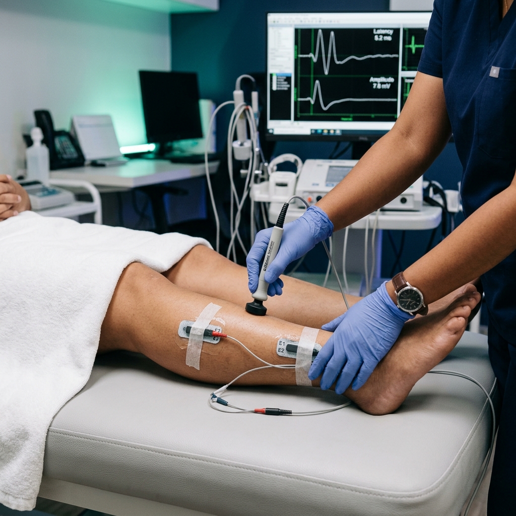
Step 2: Nerve Conduction Study (NCV)
To test for sciatica, you will lie down comfortably. Dr. Rabara will place small recording stickers on your legs and feet. Using a handheld stimulator, he will deliver mild, controlled electrical pulses to various points along your leg nerve pathways. You will feel a quick, tapping shock that lasts a fraction of a millisecond. While it may startle you slightly, it is completely safe and does not cause any lasting pain.
Step 3: Needle Electromyography (EMG)
Dr. Rabara will gently insert a microscopic, sterile needle electrode into specific muscles of your leg and lower back. This needle does not inject any medication or draw blood; it acts strictly as a microphone to listen to your muscles' natural electrical signals. You will feel a minor pinch as the needle is inserted, similar to acupuncture. You will hear a static sound on the machine's speaker, which is the sound of your muscle fibers firing. Dr. Rabara will ask you to relax your leg and then contract it slightly.
The entire test takes about 45 minutes. You can drive yourself home immediately, return to work, and resume your normal activities. Any minor, localized muscle soreness resolves completely within 24 hours.
Why a Board-Certified Physiatrist in Vigan is Your Best Diagnostic Partner
When it comes to complex spinal nerve mapping, the accuracy of your test depends entirely on the expertise of the doctor performing it.
At TeraCare Vigan, your entire EMG-NCV test is conducted from start to finish by Dr. Ben Paolo C. Rabara, a Board-Certified Physiatrist (Physical Medicine & Rehabilitation specialist).
As a pinched nerve specialist Vigan, Dr. Rabara brings unique advantages to your spinal care:
- Comprehensive Spine Mapping: Unlike standard technician-led labs, Dr. Rabara utilizes his deep medical knowledge of spinal anatomy to trace atypical nerve pathways. If he finds your leg symptoms do not match the lumbar spine MRI, he can immediately expand the test in real-time to check for nerve compression in the hip or knee in a single session.
- Integrated Spine Care: As a physiatrist, Dr. Rabara's goal is functional recovery. He does not just hand you a piece of paper with numbers. The moment your test is finished, he will explain your "Nerve Map" in detail, and immediately design a personalized, non-surgical treatment plan—whether that involves customized spine exercises, physical therapy coordination, or advanced guided spine injections.
- Advanced Local Lab: Families in Vigan City, Ilocos Sur, and Northern Luzon no longer need to travel to distant metropolitan centers like Manila just to get a high-quality nerve test. Our clinic is equipped with state-of-the-art diagnostic equipment, providing world-class accuracy right in your neighborhood, saving you from painful, expensive travel.
Conclusion: Chart Your Path to Sciatica Pain Relief
Do not let sciatica, shooting leg pain, or a pinched nerve dictate your life, and do not make critical decisions about spinal surgery based on a picture alone. A spine MRI is a vital tool, but it is only half the diagnostic equation.
A precision EMG-NCV test at TeraCare Vigan takes less than an hour, but it provides the objective, electrical proof needed to pinpoint the exact location and severity of your pinched nerve. Reclaim your mobility and get the precise answers you need to start the right treatment today.
Locate the True Source of Your Spine Pain
Stop guessing which spinal disc is causing your pain. Schedule a precision, board-certified EMG-NCV diagnostic study with Dr. Ben Rabara in Vigan City.
Clinical Review Note: This educational guide was drafted under direct physician supervision and thoroughly reviewed for electrodiagnostic accuracy, anatomical precision, and patient safety before publication.
References & Clinical Evidence
- [1] American Association of Neuromuscular & Electrodiagnostic Medicine (AANEM). (2021). Guidelines for the clinical utility of electrodiagnostic studies in cervical and lumbar radiculopathy.
- [2] Boden, S. D., et al. (1990/Re-evaluated 2022). Abnormal magnetic-resonance scans of the lumbar spine in asymptomatic subjects: A classic prospective investigation. Journal of Bone and Joint Surgery.
- [3] Cho, S. C., et al. (2020). Electrodiagnostic assessment of lumbar radiculopathy: A systematic review of diagnostic accuracy and clinical outcomes. American Journal of Physical Medicine & Rehabilitation.
- [4] North American Spine Society (NASS). (2020). Evidence-based clinical guidelines for the diagnosis and treatment of lumbar disc herniation with radiculopathy.
* Clinical references are provided to support the medical claims made in this article. TeraCare adheres to evidence-based practices in physical medicine and rehabilitation.

Dr. Ben Rabara
Dr. Ben Rabara is a Board-Certified Physiatrist specializing in Physical Medicine and Rehabilitation. He focuses on non-surgical, precision treatments for musculoskeletal conditions, utilizing advanced diagnostics like MSK Ultrasound.
Medical Disclaimer: The information provided in this article is for educational purposes only and does not substitute for professional medical advice, diagnosis, or treatment. Always consult a qualified physician for your specific health conditions.