- — Cubital Tunnel Syndrome occurs when the ulnar nerve is compressed, stretched, or irritated as it passes through a tight bony tunnel on the inside of the elbow.
- — The hallmark symptoms are numbness and tingling selectively affecting the pinky (small) finger and the adjacent half of the ring finger.
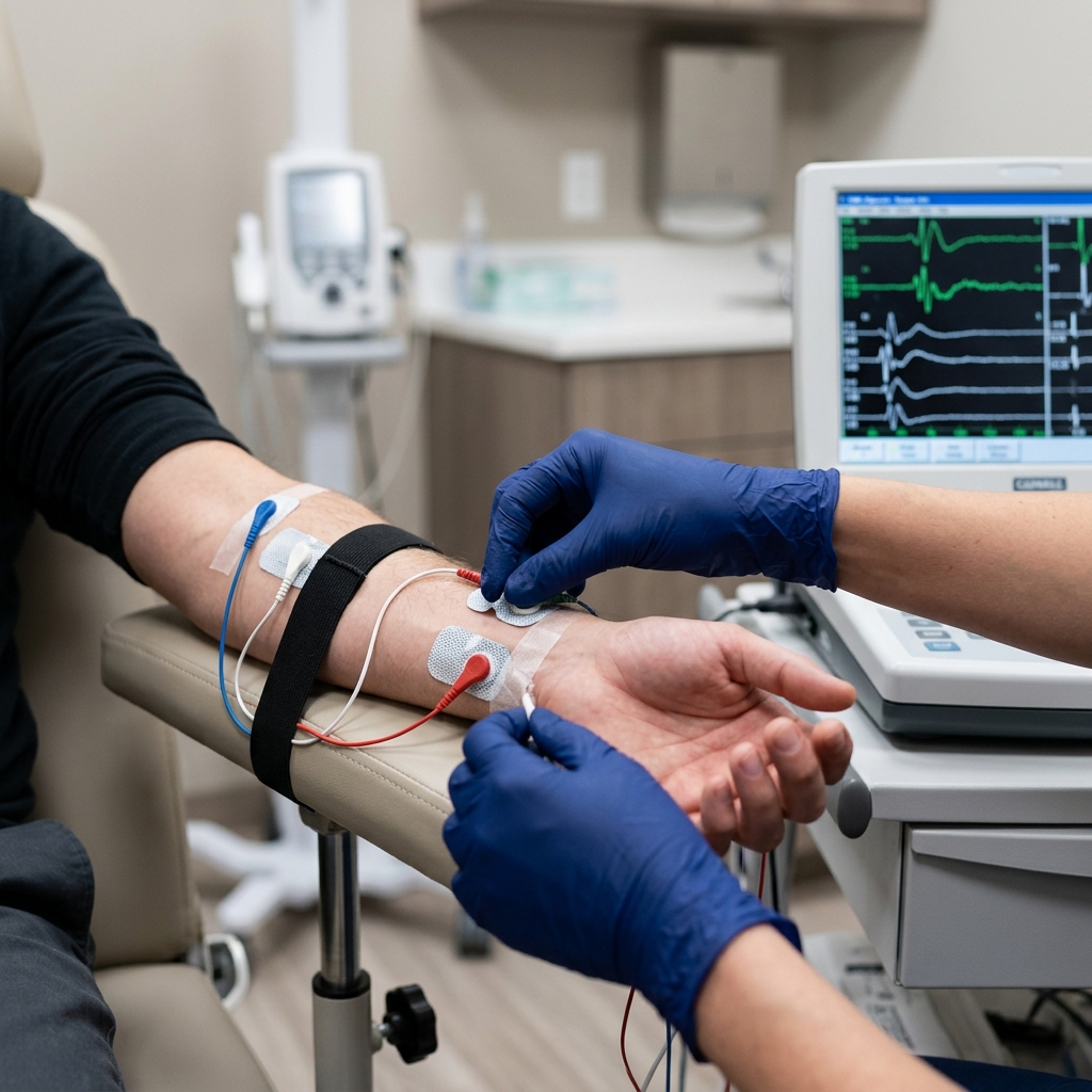
- — An NCV test mathematically isolates the compression by measuring a sudden slowdown in electrical signal velocity as it crosses the elbow crease.
- — Needle EMG evaluates the target hand muscles to rule out severe axon death and differentiate cubital tunnel from a pinched C8 nerve root in the neck.
- — Physiatrist-led conservative treatment (including nocturnal elbow splinting and specialized ulnar nerve glides) is highly effective when started early.
You wake up in the middle of the night, and your hand feels unusually cold and dead. When you shake it to bring back the feeling, you notice that the numbness is highly selective: it affects only your pinky (small) finger and the inside edge of your ring finger. During the day, you notice a sharp, electrical zap shooting down your forearm whenever you lean your elbow on a desk. When you try to type, your hand feels clumsy, and you find yourself constantly mistyping keys or struggling to spread your fingers apart.
This highly specific set of symptoms is the classic presentation of Cubital Tunnel Syndrome, which is the second most common peripheral nerve entrapment in the human body. It occurs when the ulnar nerve is mechanically compressed, stretched, or restricted in its movement as it wraps around the inner elbow joint.
Because the ulnar nerve supplies the muscles that control fine, complex hand movements, a bottleneck at the elbow can lead to rapid hand muscle wasting and permanent loss of dexterity.
To protect your hand coordination and eliminate the numbness, a prompt and highly precise EMG NCV test is an absolute clinical necessity. This functional diagnostic test takes the guesswork out of your treatment plan by proving exactly where and how severely the ulnar nerve is struggling.
The Anatomy of the Ulnar Nerve and Cubital Tunnel
The ulnar nerve is a major electrical conduit that branches off from your neck (C8 and T1 nerve roots), travels through the shoulder and down the inner side of your upper arm.
At the elbow, it must pass through a narrow bony channel called the ulnar groove (located behind the medial epicondyle, which is the hard bony bump on the inside of the elbow). Overlying this bony groove is a tight roof of fibrous tissue called Osborne's ligament. This bony channel and fibrous roof together form the cubital tunnel.
The cubital tunnel is an incredibly vulnerable anatomical area. Unlike most major nerves in the body, which are buried deep beneath protective layers of muscle and fat, the ulnar nerve in the cubital tunnel lies immediately beneath the skin. When you bump the inside of your elbow and feel a sudden, painful electric shock, you are directly striking the exposed ulnar nerve—which is why this area is colloquially called the "funny bone."
When your elbow is straight, the cubital tunnel is wide, and the ulnar nerve rests comfortably. However, when you bend your elbow:
- Tunnel Narrowing: The bending motion stretches Osborne's ligament, reducing the vertical height of the cubital tunnel and squeezing the nerve.
- Nerve Stretching: The ulnar nerve is forced to stretch tightly around the bony inner corner of the elbow, increasing its internal pressure by up to three times.
- Blood Supply Starvation: The high mechanical pressure squeezes the tiny blood vessels that feed the ulnar nerve, causing local ischemia (oxygen starvation).
If you repeatedly sleep with your elbows bent tightly, hold a phone to your ear for hours, or lean your elbows on hard surfaces while working, the constant stretching and pinching cause the nerve's protective insulation (myelin) to break down, resulting in chronic numbness and pain.
Recognizing the Progressive Stages of Ulnar Nerve Compression
Ulnar neuropathy at the elbow progresses through three clinical stages. Recognizing these symptoms early is critical to avoiding permanent hand damage:
- Mild Stage (Sensory Fluctuations): Numbness and tingling in the pinky and half of the ring finger come and go. These sensations are usually triggered by specific positions, such as driving, sleeping with bent elbows, or talking on the phone. You may feel a mild ache on the inside of your elbow.
- Moderate Stage (Persistent Sensory and Grip Loss): The numbness becomes constant, even when your arm is straight. You begin to notice a weak grip, making it difficult to open jars or carry bags. The muscles between your thumb and index finger may feel slightly soft or empty.
- Severe Stage (Axon Death & Atrophy): The ulnar nerve fibers undergo permanent structural degeneration (axonotmesis). The small muscles in your hand waste away (atrophy), creating deep hollows between your bones. You lose the ability to spread your fingers. In the most advanced cases, the ring and pinky fingers curl up tightly because the hand's natural muscle balance is lost, resulting in the permanent deformity known as an ulnar claw.
Clinical Comparison: Cubital Tunnel vs. Carpal Tunnel
Because hand numbness is so common, patients frequently confuse cubital tunnel syndrome with carpal tunnel syndrome.
However, they involve entirely different nerves, different sensory zones, and require completely different treatment plans:
| Clinical Metric | Cubital Tunnel Syndrome | Carpal Tunnel Syndrome |
|---|---|---|
| Primary Compressed Nerve | Ulnar Nerve. | Median Nerve. |
| Exact Compression Site | Inner Elbow (Cubital Tunnel). | Wrist (Carpal Tunnel). |
| Numbness Distribution | Pinky finger and the inner half of the ring finger. | Thumb, index, middle, and outer half of the ring finger. |
| Classic Physical Sign | Hollowing of the hand muscles; ulnar clawing. | Wasting of the fleshy muscle pad at the base of the thumb. |
The Gold Standard: Precision EMG-NCV for Ulnar Neuropathy
An EMG NCV test is the only diagnostic tool that can objectively prove ulnar nerve compression at the elbow. In our Vigan clinic, Dr. Ben Paolo Rabara uses a highly detailed electrodiagnostic protocol to ensure absolute accuracy:
Step 1: Segmental Nerve Conduction Studies (Inching Method)
Dr. Rabara applies recording stickers to the ulnar-innervated muscles of your hand (such as the abductor digiti minimi). He then delivers brief electrical stimulations at specific points along the arm: the wrist, just below the elbow, and just above the elbow.
By measuring the distance and transit times, he calculates the conduction velocity across the elbow crease. In a healthy ulnar nerve, signals travel faster than 50 meters per second. In cubital tunnel syndrome, we see a localized drop in speed and a reduction in signal amplitude as the current crosses the compressed elbow segment.
Step 2: Needle EMG of the Hand and Forearm
Dr. Rabara inserts a microscopic needle electrode into the small hand muscles (like the First Dorsal Interosseous) and forearm muscles (like the Flexor Carpi Ulnaris).
If the ulnar nerve's internal copper wires (axons) are actively dying, the resting hand muscles will fire abnormal spontaneous signals (fibrillations and positive sharp waves). This step is crucial because it allows us to grade the severity of the nerve damage as mild, moderate, or severe.
Step 3: Ruling out Double Crush and Spine Pathology
Symptoms of pinky numbness can also be caused by a compressed C8 spinal nerve root in your neck (cervical radiculopathy) or compression of the nerve network in your shoulder (Thoracic Outlet Syndrome). By testing non-ulnar muscles that also share the C8 pathway, Dr. Rabara can definitively rule out neck pathology, ensuring that you receive the correct treatment for your elbow.
Conservative Rehabilitation: Restoring Nerve Flow Without Surgery
Once Dr. Rabara completes your ulnar nerve map, he designs a customized, non-surgical rehabilitation plan tailored to your severity:
- Nocturnal Elbow Splinting: Because we naturally bend our elbows tightly when sleeping, wearing a lightweight nocturnal splint that holds the elbow in a comfortable, semi-straight position (around 45 degrees of flexion) is the most critical step. This immediately reduces mechanical stretch on the ulnar nerve, allowing the myelin insulation to heal overnight.
- Ulnar Nerve Gliding Exercises: Nerves need to slide smoothly through their anatomical tunnels. Our physical therapists teach you specialized ulnar nerve glides. These gentle movements slide the nerve back and forth through the cubital tunnel, breaking up inflammatory scar tissue and restoring healthy blood flow.
- Ergonomic Modifications: We help you modify your daily workspace. This includes adjusting keyboard heights to prevent sharp elbow bending, using hands-free headsets for phone calls, and utilizing specialized elbow pads to cushion the ulnar nerve from direct compression on hard desks.
- Surgical Coordination: If the needle EMG reveals severe, active axon loss that threatens permanent hand paralysis, Dr. Rabara will coordinate directly with trusted hand surgeons for a surgical decompression (releasing the tight ligament over the nerve) or ulnar nerve transposition (relocating the nerve to the front of the elbow where it is no longer stretched).
Conclusion: Protect Your Hand Coordination Today
Persistent pinky numbness, constant funny bone pain, and drop-grip clumsiness are signs that your ulnar nerve is actively losing its vital electrical current. Waiting for the symptoms to resolve on their own can lead to permanent muscle loss and irreversible hand deformities.
An EMG-NCV study is the gold-standard, functional diagnostic test that takes the guesswork out of hand pain. It proves exactly where the ulnar nerve is pinched and guides you safely back to full strength and coordination.
Don't let nerve compression steal your hand coordination. Schedule a precision, board-certified EMG-NCV study at TeraCare Vigan today.
Restore Your Hand Strength
Stop letting pinky numbness make your hand clumsy. Schedule a precision ulnar nerve study with Dr. Ben Rabara in Vigan City.
Clinical Review Note: This medical guide was written under direct physician supervision and thoroughly reviewed by Dr. Ben Paolo C. Rabara for ulnar nerve anatomical precision, electrodiagnostic accuracy, and physical rehabilitation compliance before publication.
References & Clinical Evidence
- [1] Palmer, B. A., & Hughes, T. B. (2010). Cubital Tunnel Syndrome. Journal of Hand Surgery, 35(1), 153-163.
- [2] Preston, D. C., & Shapiro, B. E. (2020). Electromyography and Neuromuscular Disorders: Clinical-Electrophysiologic Correlations (4th ed.). Elsevier.
- [3] American Association of Neuromuscular & Electrodiagnostic Medicine (AANEM). (2022). Electrodiagnostic Evaluation of Ulnar Neuropathy at the Elbow.
- [4] Cutts, S. (2007). Cubital Tunnel Syndrome. Postgraduate Medical Journal, 83(975), 28-31.
* Clinical references are provided to support the medical claims made in this article. TeraCare adheres to evidence-based practices in physical medicine and rehabilitation.

Dr. Ben Rabara
Dr. Ben Rabara is a Board-Certified Physiatrist specializing in Physical Medicine and Rehabilitation. He focuses on non-surgical, precision treatments for musculoskeletal conditions, utilizing advanced diagnostics like MSK Ultrasound.
Medical Disclaimer: The information provided in this article is for educational purposes only and does not substitute for professional medical advice, diagnosis, or treatment. Always consult a qualified physician for your specific health conditions.