- — Cervical radiculopathy occurs when a spinal nerve root in the neck becomes compressed, inflamed, or damaged, causing symptoms to shoot down the shoulder, arm, or hand.
- — An EMG-NCV study is the gold standard for differentiating a pinched nerve in the neck (C5/C6/C7/C8 roots) from distal nerve traps like Carpal Tunnel Syndrome at the wrist.
- — MRIs display physical structures (like bulging discs), but only an EMG-NCV evaluates the actual physiological health and conduction of the nerve pathway.
- — Electrodiagnostic testing provides precise localizing data, mapping the exact spinal segment that is compressed to prevent clinical misdiagnosis.
- — Early diagnosis prevents irreversible axon loss and muscle atrophy, establishing a clear roadmap for highly successful non-surgical rehabilitation.
It frequently begins as a vague, deep ache in the shoulder blade that you dismiss as a bad night's sleep. Over the next few days, however, the discomfort intensifies, transforming into a sharp, electric shock that shoots from your neck, down the side of your arm, and directly into your fingers. You might notice that tilting your head backward or turning it toward the painful side causes an unbearable surge of burning pain. Soon, a persistent numbness settles into your fingertips, and you find yourself dropping coffee mugs or struggling to hold a pen.
This debilitating constellation of symptoms is the classic presentation of cervical radiculopathy, commonly referred to as a pinched nerve in neck. It is a condition that can cause massive disruption to your daily life, making work, driving, and sleep almost impossible.
Because the nerves exiting your neck supply all the sensory pathways and muscle motor inputs of your shoulder, arm, forearm, and hand, an interruption at the spine can cause symptoms anywhere along the entire limb. This clinical overlap makes diagnosing the root cause of radiating arm pain highly complex.
Is the nerve actually compressed in your neck, or is it trapped at your elbow or wrist? To find the definitive answer, avoid guessing. A precision EMG NCV test is the absolute gold standard for mapping your nerve health and pointing the way to a non-surgical cure.
The Anatomy of Nerve Root Compression
Your cervical spine (neck) is composed of seven vertebrae (C1 through C7) stacked on top of one another. Between each vertebra is a flexible, shock-absorbing intervertebral disc. At each spinal level, a pair of spinal nerve roots branches off from the central spinal cord, exiting the bony column through tiny exit portals called neuroforamina.
These nerve roots (labeled C5, C6, C7, and C8 based on the spinal level they exit) are the main transmission cables that carry sensory feelings and motor commands between your brain and your arm. Cervical radiculopathy occurs when one of these nerve roots is mechanically compressed, chemically irritated, or severely inflamed.
There are two primary structural causes of this compression:
- Herniated or Bulging Discs: The soft, gel-like inner core of an intervertebral disc can rupture or bulge outward through a tear in its tough outer shell. This protruding disc material can directly press against the adjacent nerve root, inciting severe chemical inflammation. This is the most common cause in younger adults.
- Cervical Spondylosis (Spinal Arthritis): As we age, the spinal joints undergo natural wear and tear. The discs lose their height, and the body attempts to stabilize the spine by growing extra bone spurs (osteophytes) around the joints and neuroforamina. These hard bony growths gradually narrow the exit portals, squeezing the nerve root. This is the primary cause in older adults.
Regardless of the structural cause, the result is the same: the delicate blood supply to the nerve root is compromised, and the transmission of electrical signals is severely disrupted.
The Dermatome Map: Linking the Spine to the Hand
In my physical medicine clinic, I often show patients a dermatomal map to help them understand why a problem in their neck is felt in their fingers. A dermatome is an area of skin supplied by a single spinal nerve root.
By analyzing where your pain and numbness are located, we can make an educated clinical guess about which nerve root is struggling:
| Nerve Root | Sensory Pathway (Where You Feel It) | Motor Pathway (Muscle Weakness) | Reflex Tested |
|---|---|---|---|
| C5 Nerve Root | Outer shoulder and upper arm. | Deltoid (shoulder lift) and Biceps (elbow bend). | Biceps reflex. |
| C6 Nerve Root | Outer forearm, thumb, and index finger. | Wrist extensors (lifting hand backward). | Brachioradialis reflex. |
| C7 Nerve Root | Back of the arm, middle finger. | Triceps (elbow extension) and wrist flexors. | Triceps reflex. |
| C8 Nerve Root | Inner forearm, pinky, and ring fingers. | Hand intrinsics (finger spread, grip strength). | None (no major tendon reflex). |
While this anatomical map is extremely helpful, human biology has a significant amount of natural variation. Nerves frequently overlap, meaning a physical exam alone cannot provide the absolute, millimeter-precise localization required for surgical or injection planning.
The Diagnostic Dilemma: Pinched Neck vs. Distal Entrapments
The biggest clinical challenge in treating radiating arm pain is that distal nerve entrapment syndromes can mimic cervical radiculopathy perfectly.
For example, if you experience numbness in your thumb, index, and middle fingers, it could be caused by a compressed C6 or C7 nerve root in your neck. However, this exact same pattern is the hallmark symptom of Carpal Tunnel Syndrome, where the median nerve is compressed at the wrist. Similarly, numbness in your pinky and ring finger could be caused by a C8 nerve root pinch in your neck, or by Cubital Tunnel Syndrome, where the ulnar nerve is compressed at the elbow.
Misdiagnosing these conditions carries severe consequences. If a patient undergoes carpal tunnel release surgery at the wrist when the real problem is a compressed C6 root in their neck, their symptoms will not improve, and they will have undergone an unnecessary surgical procedure.
This is where the absolute limitation of an MRI becomes clear. An MRI is a structural photograph. It can show a bulging disc, but it cannot show if that bulging disc is actually blocking the electrical flow of the nerve. Many individuals have bulging discs on their MRIs but feel zero pain because the disc is not compressing the nerve. Conversely, a tiny, hard-to-see bone spur might be severely damaging a nerve, but it could be missed on a static scan.
An EMG NCV test is a functional study. It acts like an electrician's voltmeter, measuring the actual live electrical current flowing through your nerves and muscles. It provides clear, mathematical proof of whether a nerve is damaged, where the bottleneck is located, and how severe the compression is.
How a Precision EMG-NCV Isolates Cervical Radiculopathy
When you undergo a precision electrodiagnostic study for suspected neck issues at TeraCare Vigan, Dr. Ben Paolo Rabara personally designs and conducts a customized two-part evaluation:
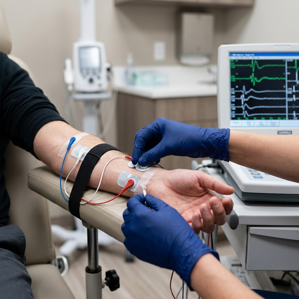
Part 1: Nerve Conduction Studies (NCS / NCV)
We apply small, metallic recording electrodes to your hand and arm. Using a handheld stimulator, we deliver a series of brief, safe electrical stimulations to test the sensory and motor branches of the median, ulnar, and radial nerves.
If the speed (conduction velocity) and signal strength (amplitude) remain perfectly normal as they pass through your wrist and elbow, we can confidently rule out localized compression like carpal tunnel or cubital tunnel syndrome.
Part 2: Needle Electromyography (EMG)
This is the most critical phase for diagnosing a pinched neck nerve. Dr. Rabara inserts a microscopic, sterile, solid-state needle electrode into specific muscles that correspond to different cervical nerve roots.
When a nerve root is pinched in the neck, the muscle fibers it feeds lose their electrical connection. Within 3 weeks, these denervated muscle fibers become highly unstable and begin firing spontaneous, abnormal electrical signals. By listening to and viewing these signals (such as fibrillation potentials and positive sharp waves), we can map the exact spinal segment that is compressed.
To confirm a cervical radiculopathy diagnosis, we look for a clear pattern: abnormal EMG findings in two or more muscles supplied by the same spinal nerve root, but controlled by different peripheral nerves. We also test your paraspinal muscles (the deep muscles immediately adjacent to your spine in the neck) to prove that the compression is occurring at the spinal exit portal rather than further down the arm.
Non-Surgical Recovery: Tailored Care Guided by Your Nerve Map
The most powerful advantage of an EMG-NCV study is that it provides a precise clinical roadmap for non-surgical recovery. In my clinical practice, we find that over 90% of cervical radiculopathy cases can be resolved completely without spine surgery:
- Mechanical Cervical Traction: Once we identify the exact compressed nerve root, we can use motorized mechanical traction to apply a gentle, therapeutic stretch to your neck. This widening of the spinal joints creates negative pressure, pulling the bulging disc material away from the nerve and instantly restoring electrical flow.
- Targeted Physical Therapy: General neck exercises can often worsen a pinched nerve. By knowing the exact nerve root involved, our physical therapists design a customized routine to strengthen the specific deep neck stabilizer muscles (like the longus colli), reducing mechanical load on the damaged spine segment.
- Precision Ultrasound-Guided Injections: If severe pain prevents you from participating in physical therapy, we can use high-resolution ultrasound to visualize your cervical spine and deliver a localized anti-inflammatory cortisone injection directly adjacent to the pinched nerve root. This calms the chemical fire, providing rapid, lasting relief.
- Posture and Ergonomic Redesign: We analyze your daily workspace, sleeping positions, and mobile phone habits (preventing 'text neck') to eliminate the repetitive mechanical strains that caused the nerve root to become trapped in the first place.
Conclusion: Reclaim Your Strength and Map Your Path to Relief
Radiculopathy is a sign that a vital pathway of your body's electrical grid is being crushed. Relying on blind physical therapy, popping pain medications continuously, or jumping straight into high-risk spine surgery based on a static MRI scan are dangerous pathways.
An EMG-NCV study is the definitive, scientifically validated test that takes the guesswork out of neck and arm pain. It shows exactly where the nerve is pinched, how severe the damage is, and how to safely heal it.
Stop suffering from radiating pain and clumsy fingers. Schedule a precision, board-certified EMG-NCV diagnostic study at TeraCare Vigan today and start your journey back to a pain-free, active life.
Stop Radiating Neck & Arm Pain
Get objective, mathematical proof of your nerve health. Schedule your board-certified EMG-NCV study with Dr. Ben Rabara in Vigan City.
Clinical Review Note: This comprehensive guide was written under direct physician supervision and thoroughly reviewed by Dr. Ben Paolo C. Rabara for physical medicine accuracy, cervical spine anatomical precision, and electrodiagnostic compliance before publication.
References & Clinical Evidence
- [1] Corey, D. L., & Comeau, D. (2014). Cervical Radiculopathy. Medical Clinics of North America, 98(4), 791-799.
- [2] Preston, D. C., & Shapiro, B. E. (2020). Electromyography and Neuromuscular Disorders: Clinical-Electrophysiologic Correlations (4th ed.). Elsevier.
- [3] American Association of Neuromuscular & Electrodiagnostic Medicine (AANEM). (2021). Guidelines for the Evaluation of Cervical Radiculopathy.
- [4] Rhee, J. M., Yoon, T., & Riew, K. D. (2007). Cervical Radiculopathy. Journal of the American Academy of Orthopaedic Surgeons, 15(8), 486-494.
* Clinical references are provided to support the medical claims made in this article. TeraCare adheres to evidence-based practices in physical medicine and rehabilitation.

Dr. Ben Rabara
Dr. Ben Rabara is a Board-Certified Physiatrist specializing in Physical Medicine and Rehabilitation. He focuses on non-surgical, precision treatments for musculoskeletal conditions, utilizing advanced diagnostics like MSK Ultrasound.
Medical Disclaimer: The information provided in this article is for educational purposes only and does not substitute for professional medical advice, diagnosis, or treatment. Always consult a qualified physician for your specific health conditions.