- — An EMG-NCV study is not a single test; it comprises two distinct diagnostic procedures: Nerve Conduction Velocity (NCV) and needle Electromyography (EMG).
- — The NCV portion uses skin stickers and mild electrical stimulations to calculate the speed and strength of signals passing through your peripheral nerves.
- — The EMG portion uses a microscopic needle electrode to 'listen' directly to the electrical activity inside your muscles at rest and during contraction.
- — This test is critical to diagnose muscle weakness causes, differentiating between nerve compression (pinched spine/wrist) and primary muscle diseases.
- — An EMG-NCV provides objective, mathematical proof of nerve damage that MRIs and standard blood tests cannot capture.
It begins as a minor inconvenience. You go to open a jar, and your grip slips. You reach to button your shirt, and your fingers feel unusually clumsy. When walking, your leg feels slightly heavy, or your toe catches on the rug. If you mention this to family or search online, the advice is highly diverse. You might be told you have "poor circulation," lack vitamins, need to stretch, or should schedule a spinal MRI.
But when you experience progressive loss of physical strength or unexplained physical fatigue, you are dealing with a breakdown in the body's electrical grid. Guessing the cause is highly dangerous. If a compressed nerve or primary muscle disease is left untreated, the fibers can undergo permanent structural degeneration.
To find the exact root cause of your physical deficits, your doctor will order a comprehensive diagnostic study. When looking for a reliable test for nerve damage, you will routinely see two terms paired together: "EMG" and "NCV."
But what is EMG test exactly, and how does it differ from an NCV?
In my clinical practice as a Physical Medicine & Rehabilitation (PM&R) specialist, I find that many patients arrive at our Vigan clinic filled with anxiety simply because they do not understand the procedure. Let us demystify this gold-standard diagnostic tool, breaking down exactly how it works, what it measures, and how it stops the guesswork to guide your recovery.
The Anatomy of the Procedure: NCV vs. Needle EMG
An electrodiagnostic study is essentially an electrician's audit of your body's wiring. Although commonly referred to as simply "an EMG," a complete study actually consists of two distinct, highly specialized tests that evaluate different parts of your neuromuscular system.
To understand the difference, think of a lamp plugged into a wall outlet. The power cord is the nerve, and the lightbulb is the muscle. If the lamp doesn't light up, you need to test both the cord's ability to carry electricity and the lightbulb's ability to convert that power into light.
Part 1: The Nerve Conduction Study (NCS / NCV)
The nerve conduction study evaluates the "power cords" of your body. It mathematically measures how quickly and strongly electrical signals travel through your peripheral nerves.
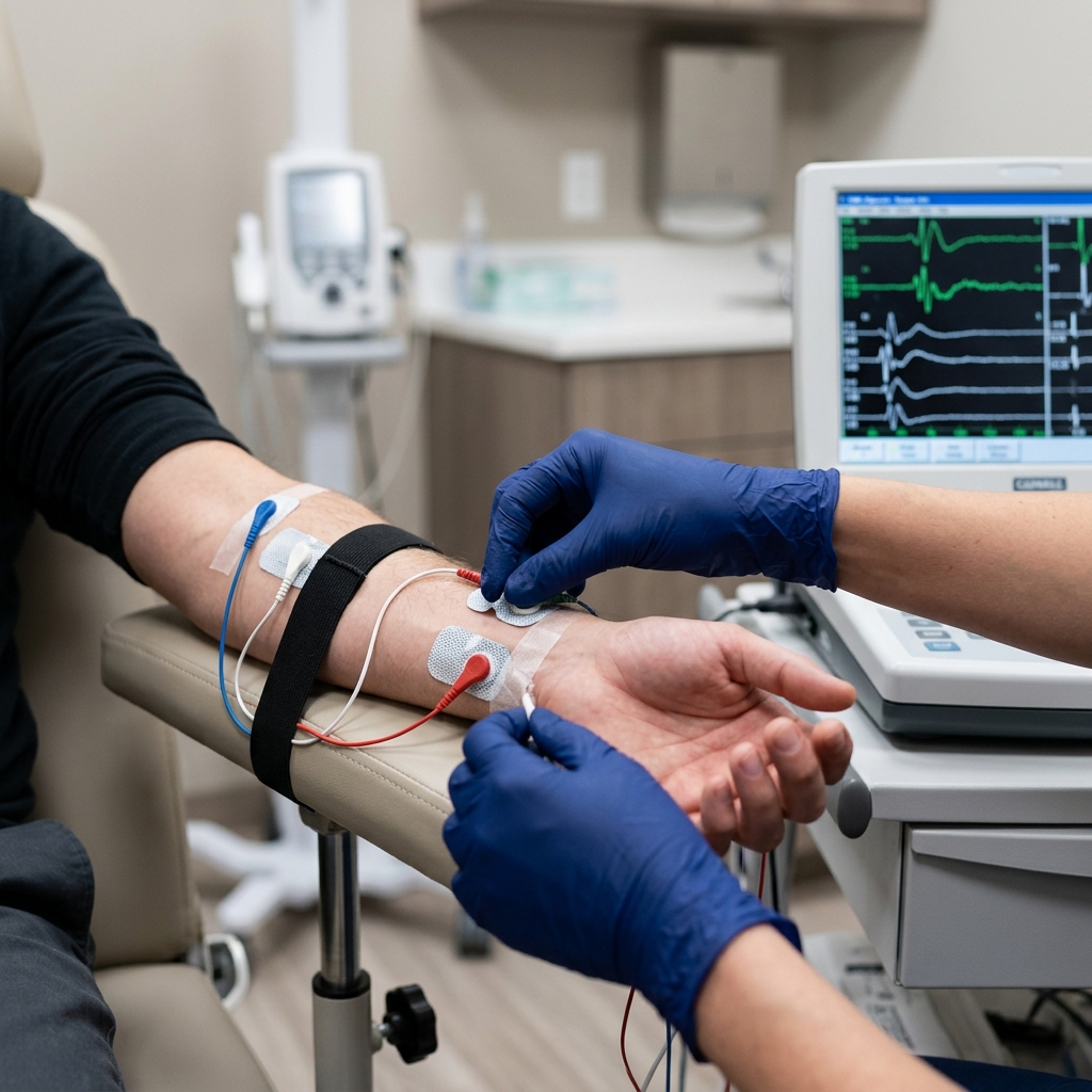
During this phase of the EMG NCV test procedure:
- Skin Stickers: We apply small, metallic recording electrodes (stickers) to the skin directly over specific nerves or the muscles they control.
- Electrical Stimulation: We use a small, handheld stimulator to deliver a brief, completely safe electrical pulse to the nerve further up the limb.
- Speed and Amplitude: Our computer records the signal as it passes. We measure two vital parameters: Conduction Velocity (how fast the signal travels, measured in meters per second) and Amplitude (the overall size of the electrical wave, showing how many nerve fibers are actively working).
If the conduction velocity is significantly slowed, it proves that the protective insulation (myelin) is damaged. If the amplitude is low, it proves that axons (the core copper wires) are actively dying.
Part 2: The Needle Electromyography (EMG)
The electromyography test evaluates the "lightbulbs"—your muscles. It analyzes the electrical environment inside the muscle tissue itself to see if the muscle is receiving healthy nerve inputs or if the muscle fibers themselves are diseased.
During this phase:
- Microscopic Needle Electrode: Dr. Rabara inserts a tiny, sterile, solid-state needle electrode into specific muscles. Note: This is not a hollow injection needle; it does not inject any medication or withdraw any blood. It acts purely as a highly sensitive microphone.
- Listening to the Muscle: The needle electrode translates the electrical activity of your muscle fibers into visual waves on a monitor and audible sounds through a speaker.
- Resting and Contracting Analysis: We analyze the muscle in two states. First, at complete rest (healthy muscles should be completely silent). Second, as you slowly contract the muscle (we listen to how the motor units recruit and fire to generate force).
By combining these two tests, we obtain a complete, three-dimensional map of your nervous and muscular health.
Diagnosing Muscle Weakness Causes: The Diagnostic Bottleneck
When patients present with physical deficits, identifying the exact source of muscle weakness causes is the most critical step in creating a successful treatment plan. Weakness is not a disease in itself; it is a symptom of a breakdown somewhere along the neuromuscular pathway.
An EMG-NCV study acts as the definitive diagnostic tool to locate the exact bottleneck:
- The Brain & Spinal Cord (Upper Motor Neuron): While the EMG-NCV primarily tests peripheral nerves, atypical results can help us rule out or suspect central nervous system issues like stroke or spinal cord injuries, prompting targeted brain imaging.
- The Spinal Nerve Roots (Radiculopathy): A herniated disc in your neck or lower back can pinch a nerve root as it exits the spine. The EMG test can trace denervation signals (like fibrillations or positive sharp waves) back to specific spinal segments (e.g., C6, L5, S1), proving the disc is actively crushing the nerve.
- The Plexus (Nerve Network): Major nerve networks in your shoulder (brachial plexus) or hip (lumbosacral plexus) can be stretched or injured. An NCV test maps these complex junctions to isolate plexopathy.
- Peripheral Nerve Entrapment (Local Compression): Nerves can be mechanically compressed at tight anatomical tunnels. This includes Carpal Tunnel Syndrome at the wrist or Cubital Tunnel Syndrome at the elbow. The NCV study mathematically proves the exact millimeter of nerve slowdown.
- Systemic Neuropathy (Metabolic/Toxic): Issues like diabetes, thyroid disease, or chronic kidney failure cause generalized nerve starvation. The NCV shows symmetrical nerve slowing, starting at the lowest extremities (feet).
- The Neuromuscular Junction: Rare diseases like Myasthenia Gravis interrupt the chemical bridge between the nerve and muscle. Specialized repetitive nerve stimulation tests can isolate this junction.
- Primary Muscle Diseases (Myopathy): Direct muscle diseases like muscular dystrophy or polymyositis present with specific small, rapid firing potentials on needle EMG, while the nerve conduction speeds remain completely normal.
Without an EMG-NCV, a doctor is forced to guess which of these seven locations is causing your weakness. Relying on blind physical therapy or undergoing spinal surgery based on a structural MRI alone carries a high clinical risk.
Clinical Comparison: NCV Study vs. Needle EMG
To help patients visualize how these two diagnostic halves collaborate, let us look at their distinct clinical functions:
| Clinical Metric | Nerve Conduction Study (NCS / NCV) | Needle Electromyography (EMG) |
|---|---|---|
| Equipment Used | Surface skin electrodes (stickers) and handheld stimulator. | Microscopic, sterile, solid-state needle electrode. |
| Primary Target | Peripheral nerve pathways (myelin and axon bundles). | Muscle fibers and active motor units. |
| Sensation Felt | Brief, millisecond tapping or twitching (static shock sensation). | A localized pinch during insertion; deep pressure during muscle contraction. |
| Primary Purpose | Assess the overall speedometer flow and size of nerve signals. | Listen for abnormal muscle firing to rule out axon death vs. primary myopathy. |
What to Expect During Your EMG-NCV Session: A Patient's Walkthrough
Knowing exactly what will happen during your appointment is the absolute best way to eliminate procedural anxiety. Here is the step-by-step clinical walkthrough at TeraCare Vigan:
Step 1: The Clinical Consultation
Before touching any equipment, Dr. Rabara will conduct a targeted history and physical exam. He will check your reflexes, muscle strength, and sensory boundaries. This allows him to customize the electrodiagnostic study, testing the exact nerves relevant to your unique symptoms rather than running a generic, incomplete template.
Step 2: Conducting the NCV
You will lie comfortably on our padded examination table. Dr. Rabara will apply the skin stickers and deliver localized electrical stimulations. While the tapping sensation can startle you initially, it is entirely tolerable. Each stimulation lasts less than a millisecond, and there are absolutely no lingering sensations or side effects.
Step 3: Conducting the Needle EMG
Dr. Rabara will clean the target muscle area. He will gently insert the sterile microscopic needle electrode into a few specific muscles (such as your hand, arm, shoulder, calf, or thigh).
You will hear a static "whooshing" or "crackling" sound from our clinical speaker, which is the actual sound of your muscle's electrical activity. Dr. Rabara will guide you to relax the muscle completely, then slowly flex it. The entire needle phase takes only 10 to 15 minutes.
Step 4: Immediate Results & Consultation
Because Dr. Rabara is a board-certified physical medicine specialist, he analyzes the electrical waveforms in real-time. You do not have to wait days for a lab technician to mail a report to another doctor. Dr. Rabara will explain the mathematical findings and direct diagnostic answers immediately after the test is completed.
The Board-Certified Physiatrist Advantage: Precision Diagnostics in Vigan City
An EMG-NCV study is not a simple automated lab test. It is a highly operator-dependent, interactive medical procedure. The accuracy of the test relies entirely on the clinical skills and physiological knowledge of the physician performing it.
This is why having your study conducted by a Board-Certified Physiatrist is an absolute necessity:
- Expert Anatomical Mapping: A physiatrist specializes in the musculoskeletal and nervous systems. Dr. Rabara knows the exact anatomical variations of peripheral nerves, ensuring that electrodes are placed with millimeter precision.
- Real-Time Diagnostic Adjustments: If Dr. Rabara detects an abnormal waveform during the NCV, he doesn't just print the error. He immediately pivots the study, testing adjacent nerve branches to isolate rare entrapments or dual pathologies (like having both carpal tunnel and a pinched neck nerve simultaneously).
- A Direct Bridge to Non-Surgical Recovery: As a PM&R specialist, Dr. Rabara's ultimate goal is functional restoration. He doesn't just hand you a diagnostic report; he translates the NCV speeds and EMG signals into a targeted physical therapy, medical bracing, or non-surgical injection plan designed to heal your specific nerve bottleneck.
- University-Grade Care in Ilocos Sur: Patients in Northern Luzon no longer need to brave the long travel, traffic, and high costs of visiting major medical centers in Metro Manila. TeraCare Vigan provides state-of-the-art diagnostic equipment and board-certified expertise right in your local community.
Conclusion: Stop Guessing and Map Your Nerve and Muscle Health Today
Unexplained muscle weakness, constant clumsiness, or heavy limbs are signs that your nervous system is actively struggling. Relying on superficial guesswork or waiting for the weakness to go away can lead to irreversible muscle atrophy and permanent nerve loss.
An EMG-NCV study is the definitive, scientifically proven test for nerve damage that provides the absolute mathematical proof your doctors need to design a successful recovery plan.
Take control of your mobility. Schedule a precision, board-certified EMG-NCV diagnostic study at TeraCare Vigan today.
Map Your Nerve and Muscle Health
Stop guessing why you have muscle weakness. Schedule a precision, board-certified EMG-NCV diagnostic study with Dr. Ben Rabara in Vigan City.
Clinical Review Note: This educational guide was drafted under direct physician supervision and thoroughly reviewed for electrodiagnostic accuracy, anatomical precision, and patient safety before publication.
References & Clinical Evidence
- [1] American Association of Neuromuscular & Electrodiagnostic Medicine (AANEM). (2022). Guidelines for Ethical Practice in Electrodiagnostic Medicine.
- [2] Preston, D. C., & Shapiro, B. E. (2020). Electromyography and Neuromuscular Disorders: Clinical-Electrophysiologic Correlations (4th ed.). Elsevier.
- [3] Katirji, B. (2021). Electromyography in Clinical Practice: A Case Study Approach (3rd ed.). Oxford University Press.
- [4] American Academy of Neurology (AAN). (2023). Clinician Summary: The Role of Electrodiagnostic Testing in Neuromuscular Disorders.
* Clinical references are provided to support the medical claims made in this article. TeraCare adheres to evidence-based practices in physical medicine and rehabilitation.

Dr. Ben Rabara
Dr. Ben Rabara is a Board-Certified Physiatrist specializing in Physical Medicine and Rehabilitation. He focuses on non-surgical, precision treatments for musculoskeletal conditions, utilizing advanced diagnostics like MSK Ultrasound.
Medical Disclaimer: The information provided in this article is for educational purposes only and does not substitute for professional medical advice, diagnosis, or treatment. Always consult a qualified physician for your specific health conditions.