- — Sports nerve injuries range from minor stretch injuries (stingers/burners) to severe peripheral compressions like common peroneal nerve damage causing foot drop.
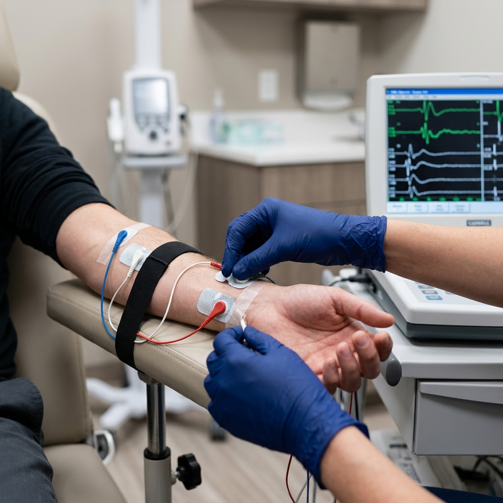
- — An EMG-NCV study is the only objective diagnostic tool that can physically differentiate temporary nerve bruising (Neuropraxia) from active fiber death (Axonotmesis).
- — A 'stinger' or 'burner' involves a traumatic traction or compression of the brachial plexus in the shoulder, requiring careful clinical mapping to ensure safe return-to-play.
- — Foot drop is commonly caused by compression of the peroneal nerve at the outer knee, which stops the signals needed to lift the foot, causing a dragging gait.
- — Serial EMG-NCV studies are highly critical to track nerve regeneration speed (typically 1 millimeter per day) and guide targeted athletic rehabilitation.
The collision is instantaneous. A hard tackle on the football field, a heavy fall on the basketball court, or an awkward twist during a sprint. In an instant, you feel a violent, white-hot flash of electricity shoot from your neck down into your fingertips, leaving your arm hanging completely limp and dead at your side. Or perhaps, after a hard impact to the outside of your knee, you stand up only to find that your foot feels unusually heavy, and you physically cannot lift your toes off the turf, forcing you to high-step like a marching soldier just to avoid tripping.
These are the alarming realities of sports nerve injuries. While athletes, coaches, and sports physical therapists are highly trained to recognize muscle tears, ligament sprains, and bone fractures, traumatic injuries to the peripheral nervous system are frequently misunderstood, neglected, or misdiagnosed.
A nerve injury is not a simple structural tear that can be verified on a standard X-ray or MRI scan. It is a functional disruption of your body's communication grid.
Whether you are dealing with a recurring shoulder stinger from contact sports or progressive foot drop after a direct leg impact, a precision EMG NCV test is the absolute gold standard for mapping the severity of the damage and establishing a safe, scientifically proven recovery pathway.
Shoulder Stingers and Burners: Brachial Plexopathy in Contact Sports
In high-impact contact sports like rugby, basketball, football, and martial arts, the shoulder and neck undergo extreme mechanical forces. The most common nerve injury resulting from these forces is a "stinger" or "burner."
A stinger is a traumatic injury to the brachial plexus—the complex web of nerves that originates in your neck and travels under your collarbone to supply the entire arm.
There are two primary mechanical ways a stinger occurs:
- Traction (Stretching) Injury: A collision forces your head to bend sharply to one side while your shoulder on the opposite side is violently pressed downward. This action stretches the brachial plexus nerves beyond their physical limits, tearing the delicate protective sheaths of the nerve fibers.
- Compression (Pinching) Injury: Your head is forced backward and to one side, compressing the spinal nerve roots as they exit the neuroforamina in the neck. This direct mechanical squeeze blocks signal transmission instantly.
The classic presentation is unmistakable: an immediate, intense burning, stinging, or electrical shock sensation shooting down one arm, accompanied by a sudden loss of muscle strength (such as being unable to lift your arm at the shoulder).
While most mild stingers resolve within minutes as signal flow returns, recurrent stingers or those with lingering weakness are signs of progressive nerve damage that requires a detailed board-certified evaluation to prevent permanent brachial plexus palsy.
Foot Drop: Common Peroneal Neuropathy at the Knee
Another highly debilitating sports injury is foot drop, which is characterized by the physical inability to lift the front part of the foot (dorsiflexion). An athlete with foot drop is forced to drag their toes along the ground or adopt a highly abnormal "steppage gait"—lifting the knee high so the foot clears the floor.
The vast majority of sports-related foot drop cases are caused by compression or trauma to the common peroneal nerve.
This nerve is a major branch of the sciatic nerve that travels down the back of the thigh and wraps tightly around the outer side of the knee, immediately below the joint line, hugging the neck of the fibula bone.
Because the peroneal nerve at the fibular neck lies immediately beneath the skin with virtually no cushioning muscle, it is incredibly vulnerable to:
- Direct Impact: A hard kick, knee-to-knee collision, or impact from a ball directly crushes the nerve against the underlying fibula bone.
- Joint Sprains and Fractures: A severe ankle sprain or a fracture of the upper fibula can stretch or tear the peroneal nerve along its pathway.
- External Compression: Repetitive wear of tight athletic braces, shin guard straps, compression boots, or tight leg wraps can cut off the microscopic blood flow to the nerve, starving it of oxygen.
The Physics of Nerve Damage: Grading Severity
To design a successful athletic recovery plan, we must first determine the exact grade of the nerve injury.
In physical medicine, we use the classic Seddon Classification to grade nerve trauma into three distinct categories:
| Nerve Injury Grade | Pathological State (What Happened) | EMG-NCV Findings | Recovery Timeline & Prognosis |
|---|---|---|---|
| 1. Neuropraxia | Temporary conduction block. The nerve's structure is fully intact, but the outer protective myelin insulation is bruised. | Local signal slowdown across the site of injury; zero active muscle denervation on needle EMG. | Excellent. Spontaneous healing occurs within days to a few weeks as the insulation repairs itself. |
| 2. Axonotmesis | Active fiber death. The internal copper wires (axons) are severed, but the surrounding protective connective tissue sleeve remains fully intact. | Severe reduction in NCV signal amplitude; active denervation signals (fibrillations) appear on needle EMG after 21 days. | Slow. The nerve must grow back from the site of injury at a rate of 1 millimeter per day (1 inch per month). Can take months. |
| 3. Neurotmesis | Complete nerve severing. Both the axons and the surrounding connective tissue sleeve are completely torn apart. | Zero electrical signal conduction; massive, permanent denervation on needle EMG. | Surgical. The nerve cannot guide its own regrowth and requires microsurgical repair or nerve grafting to restore function. |
The Role of EMG-NCV in Athlete Recovery & Return-to-Play
An EMG NCV test is the ultimate objective safety gate in sports medicine. Relying on an athlete's subjective feedback ("I feel fine") or basic manual muscle testing is highly dangerous.
An electrodiagnostic study provides three critical clinical answers:
- Isolating the Exact Compression Site: Radiating arm pain can be caused by a neck herniated disc or a shoulder stinger. Foot drop can be caused by a pinched L5 nerve root in the lower back or peroneal nerve compression at the knee. By testing the signal flow, Dr. Rabara isolates the exact millimeter of compression, preventing incorrect surgeries or ineffective rehab.
- Measuring Conduction Velocity and Axon Loss: The NCV portion calculates the precise signal speed. If the signal amplitude is preserved, it proves the injury is a mild Neuropraxia. If the amplitude is low, it proves active axon death is occurring, indicating a longer, more structured recovery period.
- Tracking Real-Time Reinnervation: Long before an athlete can physically contract their muscle or lift their foot, a needle EMG can detect microscopic electrical signals showing that the regenerating nerve has successfully re-connected to the muscle fibers. This "early warning system" allows us to safely progress physical therapy ahead of schedule.
Physiatrist-Led Sports Rehab: Our Multi-Phase Protocol in Vigan
At TeraCare Vigan, Dr. Ben Paolo Rabara combines state-of-the-art diagnostic mapping with advanced, non-surgical sports rehabilitation to return athletes safely to their peak performance:
Phase 1: Protecting the Nerve & Managing Inflammation
We immediately address the mechanical forces pinching the nerve. For foot drop, we utilize customized, lightweight Ankle-Foot Orthoses (AFO) or specialized dynamic nerve braces. These devices hold the foot in a safe, flat position, preventing ankle rolling and trips while keeping the shin muscles in a comfortable position that prevents contractures.
Phase 2: Targeted Biomechanical Re-education
Once the acute inflammation is controlled, our physical therapists use your precise EMG nerve map to design a targeted exercise program. For stingers, we focus on rebuilding deep neck stabilizers, correcting shoulder blade movement (scapular dyskinesis), and teaching protective tackling techniques. For foot drop, we use neuromuscular electrical stimulation to actively contract the shin muscles, keeping the fibers healthy while the nerve slowly regrows.
Phase 3: Serial Monitoring & Return-to-Play Decisions
We perform follow-up electrodiagnostic studies at highly structured intervals (typically every 6 to 8 weeks) to measure the exact speed of nerve regrowth. Dr. Rabara uses these objective mathematical metrics to make the final, safe return-to-play decision, protecting the athlete from recurrent, career-ending nerve trauma.
Conclusion: Stop Guessing and Map Your Athletic Recovery Today
Ignoring a shoulder stinger or attempting to run through foot drop are dangerous decisions that can result in permanent muscle wasting, chronic joint instability, and irreversible nerve loss.
An EMG-NCV study is the definitive, scientifically validated diagnostic test that removes the guesswork from sports medicine. It grades the severity of your injury, locates the exact site of compression, and guides your recovery safely back to the competitive arena.
Take control of your athletic career. Schedule a precision, board-certified EMG-NCV diagnostic study at TeraCare Vigan today.
Reclaim Your Athletic Performance
Stop struggling with radiating nerve pain or foot drop. Schedule a precision sports EMG-NCV study with Dr. Ben Rabara in Vigan City.
Clinical Review Note: This specialized sports medicine guide was drafted under direct physician supervision and thoroughly reviewed by Dr. Ben Paolo C. Rabara for athletic nerve injury precision, peroneal neuropathy diagnostics, and physical rehabilitation compliance before publication.
References & Clinical Evidence
- [1] Weinberg, J., & Rhee, J. M. (2010). Cervical Spine Stingers and Burners in Athletes. Current Sports Medicine Reports, 9(1), 35-41.
- [2] Preston, D. C., & Shapiro, B. E. (2020). Electromyography and Neuromuscular Disorders: Clinical-Electrophysiologic Correlations (4th ed.). Elsevier.
- [3] Poage, C., Roth, C., & Grantham, B. (2016). Peroneal Nerve Palsy: Evaluation and Management. Journal of the American Academy of Orthopaedic Surgeons, 24(1), 1-10.
- [4] American Academy of Neurology (AAN). (2022). Neurodiagnostic Evaluation of Traumatic Nerve Lesions in Sports Medicine.
* Clinical references are provided to support the medical claims made in this article. TeraCare adheres to evidence-based practices in physical medicine and rehabilitation.

Dr. Ben Rabara
Dr. Ben Rabara is a Board-Certified Physiatrist specializing in Physical Medicine and Rehabilitation. He focuses on non-surgical, precision treatments for musculoskeletal conditions, utilizing advanced diagnostics like MSK Ultrasound.
Medical Disclaimer: The information provided in this article is for educational purposes only and does not substitute for professional medical advice, diagnosis, or treatment. Always consult a qualified physician for your specific health conditions.