Explore All Joint Aspirations

Gout Flare-Ups & Rheumatoid Arthritis:
Managing Severe Joint Swelling

Discover how mechanical joint aspiration and intra-articular anti-inflammatory therapy arrest acute crystal synovitis, relieve pressure, and protect joint cartilage.

By: Dr. Ben Rabara Updated:
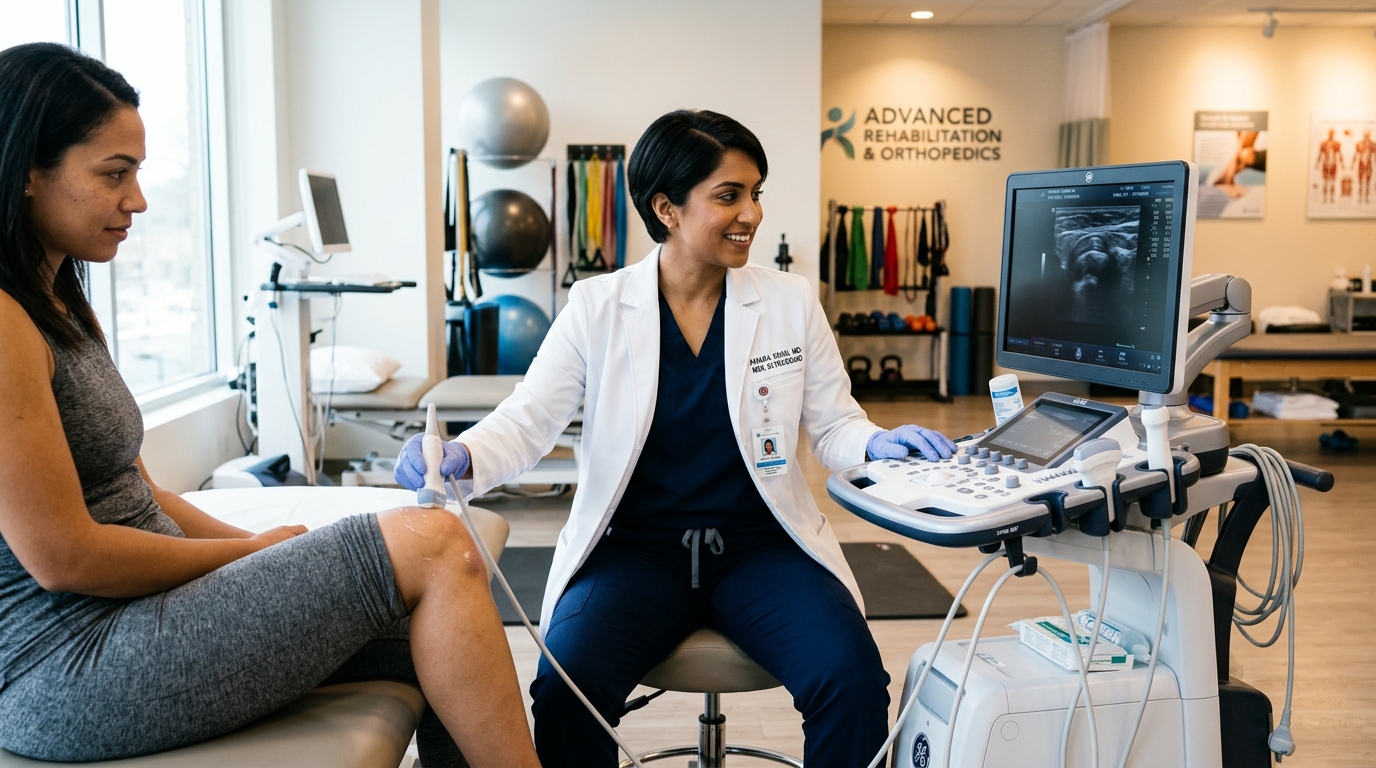
Physiatrist performing joint aspiration and injection for severe gout swelling in Vigan clinic
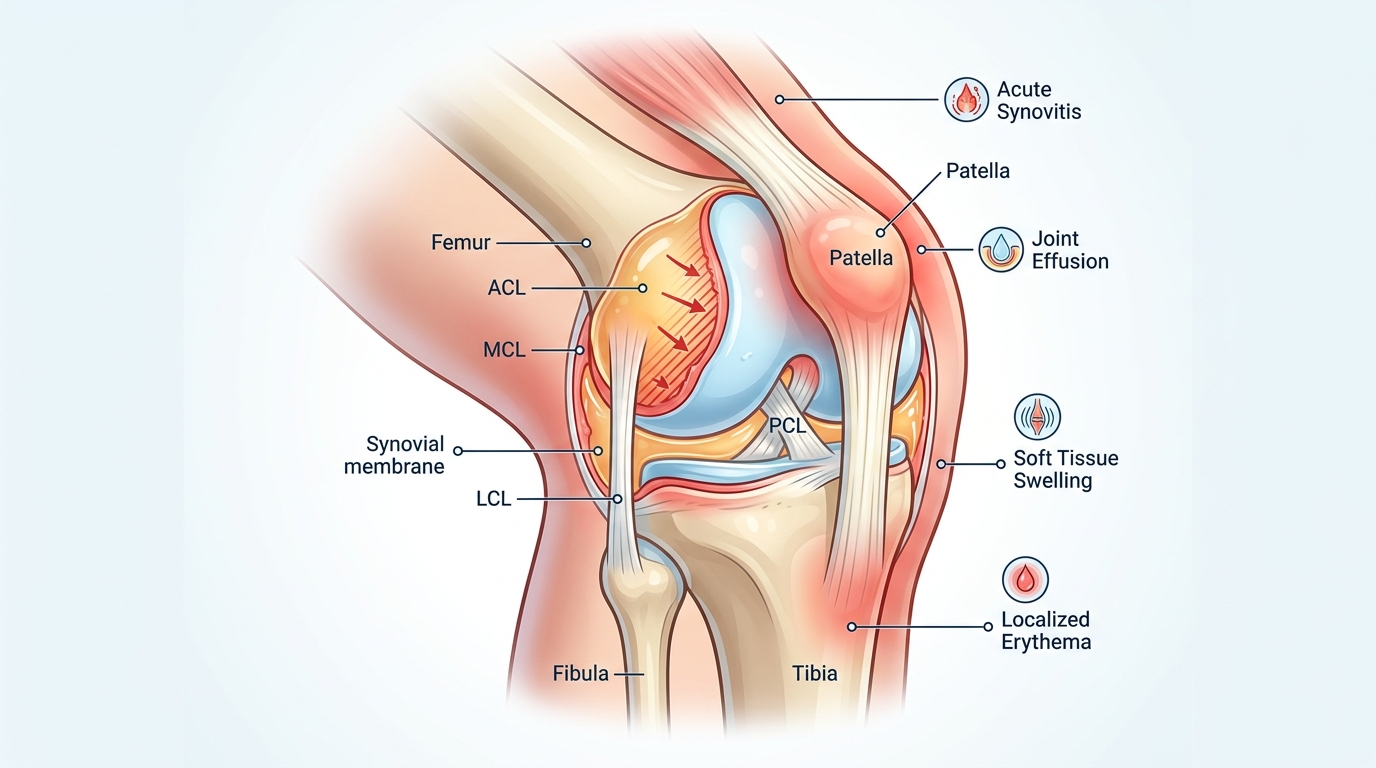
Physiatrist performing joint aspiration and injection for severe gout swelling in Vigan clinic — TeraCare Clinic Medical Illustration
Summary / Key Takeaways
  • Acute gout flare-ups precipitate monosodium urate (MSU) micro-crystals into the joint space, triggering massive neutrophil accumulation and intra-articular pressure exceeding 50 mmHg.
  • Rheumatoid arthritis (RA) flares generate an autoimmune cytokine storm (IL-1, TNF-alpha) and synovial pannus proliferation, resulting in severe joint effusion and morning stiffness lasting over 60 minutes.
  • High intra-articular pressure (>50 mmHg) collapses synovial capillary blood flow, creating ischemic cartilage hypoxia and accelerating permanent joint destruction if left undrained.
  • Therapeutic joint aspiration provides immediate mechanical decompression, followed by targeted intra-articular corticosteroid administration (Triamcinolone 40 mg) to arrest acute inflammatory synovitis.

An acute gout attack or rheumatoid arthritis (RA) flare-up is one of the most painful conditions in clinical medicine. Within hours, a joint can transform from normal into a red, hot, severely swollen structure under intense internal pressure. While oral anti-inflammatory medications play an important long-term role, they work systemically and take time to act. When a joint is distended with 20 mL to 60 mL of toxic inflammatory fluid, therapeutic joint aspiration combined with targeted intra-articular therapy provides the fastest path to immediate pain relief and joint preservation.

In my clinic, I evaluate patients presenting with acute inflammatory joint flares every week. When a patient arrives with a stiff, heavy, and catch-prone joint accompanied by a sharp pain, dull ache, radiating discomfort, burning sensation, weak feeling, tingling sensation, or grinding feeling in the knee, I look for objective signs of capsule distension. On patient forums like Reddit and medical support groups, patients suffering from gout flares frequently describe the sensation of an "internal pressure cooker about to burst." In my practice, I recommend immediate therapeutic joint aspiration to evacuate high-pressure fluid, remove abrasive micro-crystals, and arrest acute synovitis. Below, we examine the distinct pathophysiology of gout vs. RA effusions, the danger of high-pressure cartilage ischemia, and interventional treatment protocols.

Pathophysiology: Gout Micro-Crystals vs. Rheumatoid Cytokine Storms

Severe joint swelling is driven by different inflammatory mechanisms depending on the underlying pathology:

Condition Inflammatory Trigger & Mechanism Effusion Characteristics Therapeutic Aspiration Goal
Acute Gout Flare-Up Monosodium urate (MSU) micro-crystals; NLRP3 inflammasome activation High volume (20 mL – 60 mL+), cloudy, intense pressure (>50 mmHg) Evacuate abrasive micro-crystals, drop pressure, administer steroid
Rheumatoid Arthritis Flare Autoimmune pannus proliferation; TNF-alpha, IL-1 cytokine storm Translucent to cloudy yellow fluid, morning stiffness >60 mins Decompress joint, remove toxic cytokine fluid, arrest synovitis
Knee Bursitis Prepatellar / infrapatellar bursa friction or micro-trauma Localized fluid sac on top of kneecap, superficial tenderness Drain bursa fluid, relieve local friction, apply compression
Septic Arthritis (Emergency) Bacterial infection (Staph aureus); severe purulent reaction Opaque, purulent fluid; WBC >50,000 cells/mcL (>85% PMNs) Emergency drainage, urgent Gram stain, immediate IV antibiotics
Table: Clinical Comparison of Acute Inflammatory Joint Swelling Etiologies
  • Acute Gout Flare-Ups (Monosodium Urate Synovitis): Gout occurs when hyperuricemia causes monosodium urate (MSU) crystals to precipitate into the joint cavity. These microscopic, needle-shaped crystals trigger the NLRP3 inflammasome pathway in synovial macrophages, initiating an explosive inflammatory cascade. Neutrophils flood the joint space, releasing proteolytic enzymes and producing severe, rapid fluid accumulation.
  • Rheumatoid Arthritis (RA) Flares (Autoimmune Synovitis): In rheumatoid arthritis, an autoimmune reaction drives synovial lining hypertrophy (pannus formation). Pro-inflammatory cytokines—specifically Tumor Necrosis Factor-alpha (TNF-alpha), Interleukin-1 (IL-1), and Interleukin-6 (IL-6)—stimulate synovial hypersecretion, resulting in high-volume effusions, marked morning stiffness (often exceeding 60 minutes), and joint warmth.
  • Prepatellar & Infrapatellar Bursitis: Mechanical friction (e.g., prolonged kneeling) or localized trauma causes inflammation of the protective bursa sacs surrounding the kneecap. The bursa distends with inflammatory fluid, creating a distinct, superficial swelling separate from the main joint cavity.

Intra-Articular Pressure & Ischemic Cartilage Protection

When fluid accumulates rapidly inside a closed joint capsule, intra-articular pressure rises dramatically. Under resting conditions, normal knee joint pressure is slightly negative or close to 0 mmHg. During a severe gout or RA effusion, capsule pressure can surge past 50 mmHg.

This elevated hydrostatic pressure creates serious vascular consequences:

  • Synovial Capillary Occlusion: When intra-articular pressure exceeds subchondral capillary perfusion pressure (typically ~30–40 mmHg), blood flow through the synovial microvasculature collapses.
  • Chondrocyte Ischemia & Hypoxia: Articular cartilage relies on synovial fluid micro-circulation for oxygen and nutrient delivery. Prolonged pressure-induced ischemia starves chondrocytes, leading to accelerated cartilage breakdown and irreversible joint space narrowing.
  • Immediate Decompression Benefit: Evacuating 30 mL to 60 mL of fluid via therapeutic joint aspiration immediately lowers intra-articular pressure back below 10 mmHg, restoring microvascular perfusion within 15 minutes.

The Critical Rule-Out: Gout vs. Septic Arthritis Emergency

Before injecting any corticosteroid medication into an acutely swollen joint, a strict clinical rule-out protocol must be enforced. Septic arthritis—a bacterial infection of the joint space—can present with symptoms identical to a severe gout attack: intense heat, redness, extreme tenderness, and massive effusion.

Administering a steroid injection into an undiagnosed septic joint can cause catastrophic bacterial proliferation. Therefore, therapeutic aspiration serves a vital dual role: it decompressing the joint while allowing immediate diagnostic evaluation of the fluid clarity, WBC count, and Gram stain to rule out infection definitively before anti-inflammatory therapy is administered.

Therapeutic Aspiration & Targeted Intra-Articular Therapy Protocol

Once a septic joint is ruled out, our interventional protocol for acute gout and RA flares follows a dual-action approach:

  • Step 1: Complete Fluid Evacuation: Under real-time Point-of-Care MSK Ultrasound (POCUS) guidance, the aspirating needle is positioned precisely in the suprapatellar pouch. All accessible inflammatory fluid and floating crystal debris are evacuated, decompressing the joint capsule completely.
  • Step 2: Targeted Intra-Articular Steroid Administration: Without removing the needle sheath, a pre-loaded syringe containing 40 mg of Triamcinolone Acetonide (or equivalent corticosteroid) combined with local anesthetic is injected directly into the empty joint space.
  • Step 3: Sustained Anti-Inflammatory Control: While fluid removal provides instant mechanical relief, the intra-articular steroid suppresses local cytokine production at the synovial membrane, providing sustained anti-inflammatory protection for 4 to 12 weeks.

Expert Inflammatory Joint Care in Vigan City

At TeraCare Physical Medicine & Rehabilitation Clinic in Vigan City, Dr. Ben Rabara provides specialized interventional evaluation, diagnostic fluid analysis, and ultrasound-guided joint treatments for patients suffering from acute gout attacks, rheumatoid arthritis flares, and severe bursitis.

For deeper diagnostic details on ultrasound needle tracking, read our Ultrasound Aspiration & Synovial Fluid Analysis Guide. For step-by-step procedure details and local numbing protocols, consult our Knee Aspiration Procedure Deep-Dive.

Schedule Your Inflammatory Joint Evaluation

Get fast relief from severe gout flares or rheumatoid joint swelling. Book an expert evaluation with Dr. Ben Rabara in Vigan City today.

Official Medical Transparency Protocol

Clinical Realities of Inflammatory Joint Swelling

Understanding crystal synovitis, cytokine storms, and high-pressure joint preservation.

Immediate Pressure Decompression

Evacuating 20 mL to 60 mL of inflammatory fluid instantly relieves painful capsule tension and stops ischemic capillary collapse.

Removal of Destructive Micro-Crystals

Aspiration removes physical MSU gout crystals and inflammatory cytokines directly from the joint cavity.

Infection Rule-Out Safety Standard

Diagnostic fluid evaluation ensures septic arthritis is definitively excluded before administering intra-articular corticosteroids.

References & Clinical Evidence

  • [1] Elsawy AM, et al. Diagnostic accuracy of point-of-care ultrasound versus clinical physical examination for detection of knee joint effusion. BMC Musculoskelet Disord. 2023. PMID: 10257372.
  • [2] Dainese P, Wyngaert KV, De Mits S, Wittoek R, Van Ginckel A, Calders P. Association between knee inflammation and knee pain in patients with knee osteoarthritis: a systematic review. Osteoarthritis Cartilage. 2022 Apr;30(4):516-534. doi: 10.1016/j.joca.2021.12.003. PMID: 34968719.
  • [3] Sukerkar PA, Doyle Z. Imaging of Osteoarthritis of the Knee. Radiol Clin North Am. 2022 Jul;60(4):583-596. doi: 10.1016/j.rcl.2022.03.004. PMID: 35672093.
  • [4] Aiyer R, et al. Therapeutic Ultrasound for Chronic Pain Management in Joints: A Systematic Review. Pain Med. 2020. PMID: 31095336.

* Clinical references are provided to support the medical claims made in this article. TeraCare adheres to evidence-based practices in physical medicine and rehabilitation.

Dr. Ben Rabara
Medical Reviewer & Author

Dr. Ben Rabara

Dr. Ben Rabara is a Board-Certified Physiatrist specializing in Physical Medicine and Rehabilitation. He focuses on non-surgical, precision treatments for musculoskeletal conditions, utilizing advanced diagnostics like MSK Ultrasound.

Medical Disclaimer: The information provided in this article is for educational purposes only and does not substitute for professional medical advice, diagnosis, or treatment. Always consult a qualified physician for your specific health conditions.

Patient Clarity

Common Questions

Why does an acute gout attack cause such severe joint swelling?

During a gout flare, monosodium urate (MSU) crystals precipitate into the joint space. This triggers an intense NLRP3 inflammasome reaction, flooding the joint capsule with inflammatory fluid (20 mL to 60 mL or more) and raising intra-articular pressure rapidly.

How does joint aspiration help a rheumatoid arthritis flare-up?

In rheumatoid arthritis, an acute flare produces excess synovial fluid loaded with pro-inflammatory cytokines (TNF-alpha, IL-1). Evacuating this toxic fluid immediately reduces capsule distension, protects cartilage from enzymatic destruction, and prepares the joint for local anti-inflammatory therapy.

What is knee bursitis swelling?

Knee bursitis is inflammation of a fluid-filled bursa sac—most commonly the prepatellar bursa (front of the kneecap) or infrapatellar bursa. Mechanical friction or micro-trauma causes the bursa to fill with fluid, creating a distinct, tender lump on top of the joint.

Can an intra-articular steroid injection be given during an acute gout flare?

Yes, but only after therapeutic joint aspiration has successfully evacuated the excess fluid and a septic joint infection has been clinically ruled out. Administering 40 mg of triamcinolone acetonide into a decompressed joint provides powerful, sustained local anti-inflammatory control.

How do specialists tell acute gout apart from a septic joint emergency?

A septic joint is a medical emergency caused by bacterial infection. We evaluate fluid clarity, Gram stain, and white blood cell counts. Septic effusions typically present with opaque, purulent fluid and WBC counts exceeding 50,000 to 100,000 cells/mcL (>85% PMNs).

Does draining a gouty joint prevent permanent joint damage?

Yes. Prolonged high intra-articular pressure (>50 mmHg) restricts capillary blood flow to articular cartilage, leading to chondrocyte ischemia. Evacuating the effusion immediately restores microvascular perfusion and preserves joint architecture.

How long does relief last after joint aspiration and injection?

Patients experience immediate mechanical relief the moment high-pressure fluid is evacuated (typically within 15 minutes). The co-administered intra-articular corticosteroid takes effect over 24 to 48 hours and provides 4 to 12 weeks of sustained anti-inflammatory control.

Where can I get expert treatment for gout or RA joint swelling in Vigan City?

Dr. Ben Rabara provides specialized interventional MSK evaluations, joint aspirations, and targeted intra-articular therapy at TeraCare Physical Medicine & Rehabilitation Clinic in Vigan City, Ilocos Sur.

Ready to extinguish the pain?

Schedule a high-precision, ultrasound-guided evaluation with Dr. Rabara.

Message Us to Book
Chat with us