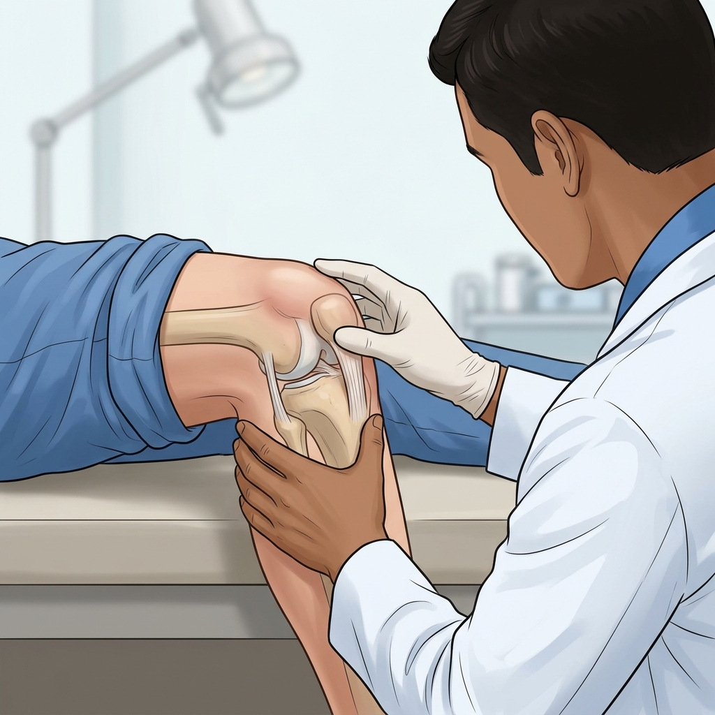
- — Point-of-Care MSK Ultrasound (POCUS) achieves over 95% diagnostic sensitivity, detecting joint effusions as small as 0.5 mL to 2.0 mL that physical palpation frequently misses.
- — Real-time in-plane needle visualization tracks the bright echogenic tip directly into the suprapatellar bursa, completely eliminating blind needle guesswork and protecting articular cartilage.
- — Laboratory synovial fluid classification differentiates 4 distinct clinical groups: Group I (Non-inflammatory), Group II (Inflammatory Gout/RA), Group III (Septic Emergency >50k WBC), and Group IV (Hemorrhagic/Fracture).
- — Compensated Polarized Light Microscopy (CPLM) identifies pathognomonic crystal structures—distinguishing needle-shaped Monosodium Urate (MSU gout) crystals from rhomboid Calcium Pyrophosphate (CPPD pseudogout) crystals.
Accurately diagnosing and treating a swollen joint requires moving beyond guesswork. For decades, traditional joint fluid removal relied on "blind" anatomical landmark palpation—a technique associated with a 20% to 30% failure rate, painful needle repositioning, and risk of cartilage irritation. Modern interventional physical medicine and rehabilitation has transformed this procedure through ultrasound-guided joint aspiration and high-complexity synovial fluid analysis.
In my clinic, I look for objective sonographic markers before placing any diagnostic needle. When evaluating patients presenting with a stiff, heavy, and catch-prone joint accompanied by a sharp pain, dull ache, radiating discomfort, burning sensation, weak feeling, tingling sensation, or grinding feeling in the knee, I rely on Point-of-Care MSK Ultrasound (POCUS) to visualize fluid pockets down to 0.5 mL in volume. On patient community forums like Reddit and medical discussion groups, educated patients increasingly seek out specialists who utilize real-time imaging rather than blind punctures. In my practice, I recommend pairing real-time ultrasound needle guidance with rigorous laboratory fluid analysis to deliver unmatched clinical safety and diagnostic clarity. Below, we examine the sonographic physics of POCUS needle tracking, the 4-group synovial fluid diagnostic classification system, and polarized crystal microscopy.
The Science of Point-of-Care MSK Ultrasound (POCUS) Guidance
Point-of-Care MSK Ultrasound (POCUS) utilizes high-frequency linear transducers (typically operating between 8 MHz and 15 MHz) to produce real-time, high-resolution cross-sectional images of joint soft tissues. POCUS achieves an extraordinary diagnostic sensitivity exceeding 95% for detecting suprapatellar bursa effusions.
Unlike blind landmark punctures, POCUS guidance offers distinct sonographic advantages:
- Quantitative Effusion Thresholds: In a healthy knee, the suprapatellar pouch contains a fluid stripe measuring less than 2 mm. An ultrasound measurement showing a fluid thickness of 4 mm or greater confirms a clinically significant joint effusion.
- Real-Time In-Plane Needle Visualization: Using an in-plane ultrasound approach, the entire length of the sterile aspirating needle—including the bright, echogenic needle tip—is monitored continuously as it slides into the fluid pocket. This prevents accidental contact with sensitive articular cartilage or periosteal bone.
- Sonographic Signs & Pathognomonic Identifiers: POCUS imaging identifies specific disease markers on the monitor, including the "Double Contour Sign" (pathognomonic for monosodium urate crystal deposition on articular cartilage in gout), the "Snowstorm appearance" (floating hyperechoic synovial debris), and Power Doppler hypervascularity indicating active acute synovitis.
The 4-Group Synovial Fluid Laboratory Classification System
The synovial fluid evacuated during an ultrasound-guided aspiration serves as a crucial diagnostic specimen. In laboratory pathology, evacuated joint fluid is systematically categorized into four distinct diagnostic groups based on physical clarity, viscosity, white blood cell (WBC) count, and differential neutrophil percentage:
| Synovial Fluid Group | Physical Appearance | WBC Count (cells/mcL) | Neutrophils (% PMN) | Primary Clinical Etiologies |
|---|---|---|---|---|
| Group I: Non-Inflammatory | Clear to light straw-yellow; High viscosity | < 2,000 | < 25% | Osteoarthritis, traumatic irritation, meniscal degenerative tears |
| Group II: Inflammatory | Translucent to cloudy yellow; Decreased viscosity | 2,000 – 50,000 | 50% – 75% | Acute Gout, Pseudogout (CPPD), Rheumatoid Arthritis, Psoriatic Arthritis |
| Group III: Septic (Emergency) | Opaque, turbid, or purulent; Very low viscosity | > 50,000 – 100,000+ | > 85% – 90% | Bacterial Septic Arthritis (Staphylococcus aureus, Neisseria gonorrhoeae) |
| Group IV: Hemorrhagic | Red, bloody, or xanthochromic; Variable viscosity | RBCs predominant | Variable | Acute ACL ligament tear, patellar dislocation, Lipohemarthrosis (fracture) |
Compensated Polarized Light Microscopy: Gout vs. Pseudogout
When a Group II inflammatory effusion is identified, differentiating between acute gout and pseudogout is essential for targeted therapy. In our diagnostic workflow, we utilize Compensated Polarized Light Microscopy (CPLM) equipped with a 550 nm red compensator plate to examine wet synovial fluid preparations:
- Monosodium Urate (MSU) Crystals (Gout): Under polarized light, MSU crystals appear as sharp, needle-shaped structures that exhibit strong negative birefringence. When aligned parallel to the slow axis of the red compensator plate, MSU crystals refract bright yellow light; when aligned perpendicular, they refract blue light.
- Calcium Pyrophosphate Dihydrate (CPPD) Crystals (Pseudogout): CPPD crystals appear as small, rhomboid or parallelepiped structures exhibiting weak positive birefringence. When aligned parallel to the compensator axis, CPPD crystals refract weak blue light; when aligned perpendicular, they refract yellow light.
Identifying Lipohemarthrosis: The Parfait Sign of Occult Fractures
When an aspiration yields frankly bloody joint fluid (Group IV Hemorrhagic), careful diagnostic inspection is required. If the evacuated syringe is allowed to stand upright for 10 to 15 minutes, or if an ultrasound scan is performed in a resting position, a three-layer fluid separation—known clinically as the "Parfait Sign" or lipohemarthrosis—may become visible.
Lipohemarthrosis occurs when an intra-articular fracture breaks the subchondral bone cortex, allowing marrow fat droplets to leak into the joint cavity alongside blood. On ultrasound and standing fluid inspection, the yellow fat layer floats on top of the dark red blood. Identifying lipohemarthrosis provides immediate diagnostic proof of an occult intra-articular bone fracture, prompting urgent radiographic CT or MRI imaging.
Diagnostic Precision & Clinical Consultation in Vigan City
At TeraCare Physical Medicine & Rehabilitation Clinic in Vigan City, Dr. Ben Rabara utilizes advanced Point-of-Care MSK Ultrasound (POCUS) to perform safe, precise joint aspirations and comprehensive diagnostic fluid evaluations.
For step-by-step details on procedure comfort and local lidocaine numbing, read our Knee Aspiration Procedure Deep-Dive. For full outpatient pricing and PhilHealth RVS 20610 coverage details, consult our Knee Aspiration Cost Guide.
Schedule Your Diagnostic POCUS Evaluation
Get sub-millimeter ultrasound accuracy and expert diagnostic fluid analysis. Book your consultation with Dr. Ben Rabara in Vigan City today.
Clinical & Sonographic Realities of Joint Evaluation
Understanding ultrasound visualization, sub-millimeter precision, and diagnostic fluid science.
Sub-Millimeter POCUS Needle Tracking
Real-time high-frequency ultrasound displays the exact depth of the fluid pocket and tracks the needle tip safely away from hyaline cartilage.
Microscopic Crystal Differentiation
Polarized light microscopy identifies whether joint swelling is driven by needle-shaped MSU gout crystals or rhomboid CPPD pseudogout crystals.
Infection Rule-Out Protocol
Synovial fluid WBC counts and Gram stains provide immediate objective proof to rule out life-threatening septic arthritis emergencies.
References & Clinical Evidence
- [1] Elsawy AM, et al. Diagnostic accuracy of point-of-care ultrasound versus clinical physical examination for detection of knee joint effusion. BMC Musculoskelet Disord. 2023. PMID: 10257372.
- [2] Dainese P, Wyngaert KV, De Mits S, Wittoek R, Van Ginckel A, Calders P. Association between knee inflammation and knee pain in patients with knee osteoarthritis: a systematic review. Osteoarthritis Cartilage. 2022 Apr;30(4):516-534. doi: 10.1016/j.joca.2021.12.003. PMID: 34968719.
- [3] Sukerkar PA, Doyle Z. Imaging of Osteoarthritis of the Knee. Radiol Clin North Am. 2022 Jul;60(4):583-596. doi: 10.1016/j.rcl.2022.03.004. PMID: 35672093.
- [4] Aiyer R, et al. Therapeutic Ultrasound for Chronic Pain Management in Joints: A Systematic Review. Pain Med. 2020. PMID: 31095336.
* Clinical references are provided to support the medical claims made in this article. TeraCare adheres to evidence-based practices in physical medicine and rehabilitation.

Dr. Ben Rabara
Dr. Ben Rabara is a Board-Certified Physiatrist specializing in Physical Medicine and Rehabilitation. He focuses on non-surgical, precision treatments for musculoskeletal conditions, utilizing advanced diagnostics like MSK Ultrasound.
Medical Disclaimer: The information provided in this article is for educational purposes only and does not substitute for professional medical advice, diagnosis, or treatment. Always consult a qualified physician for your specific health conditions.