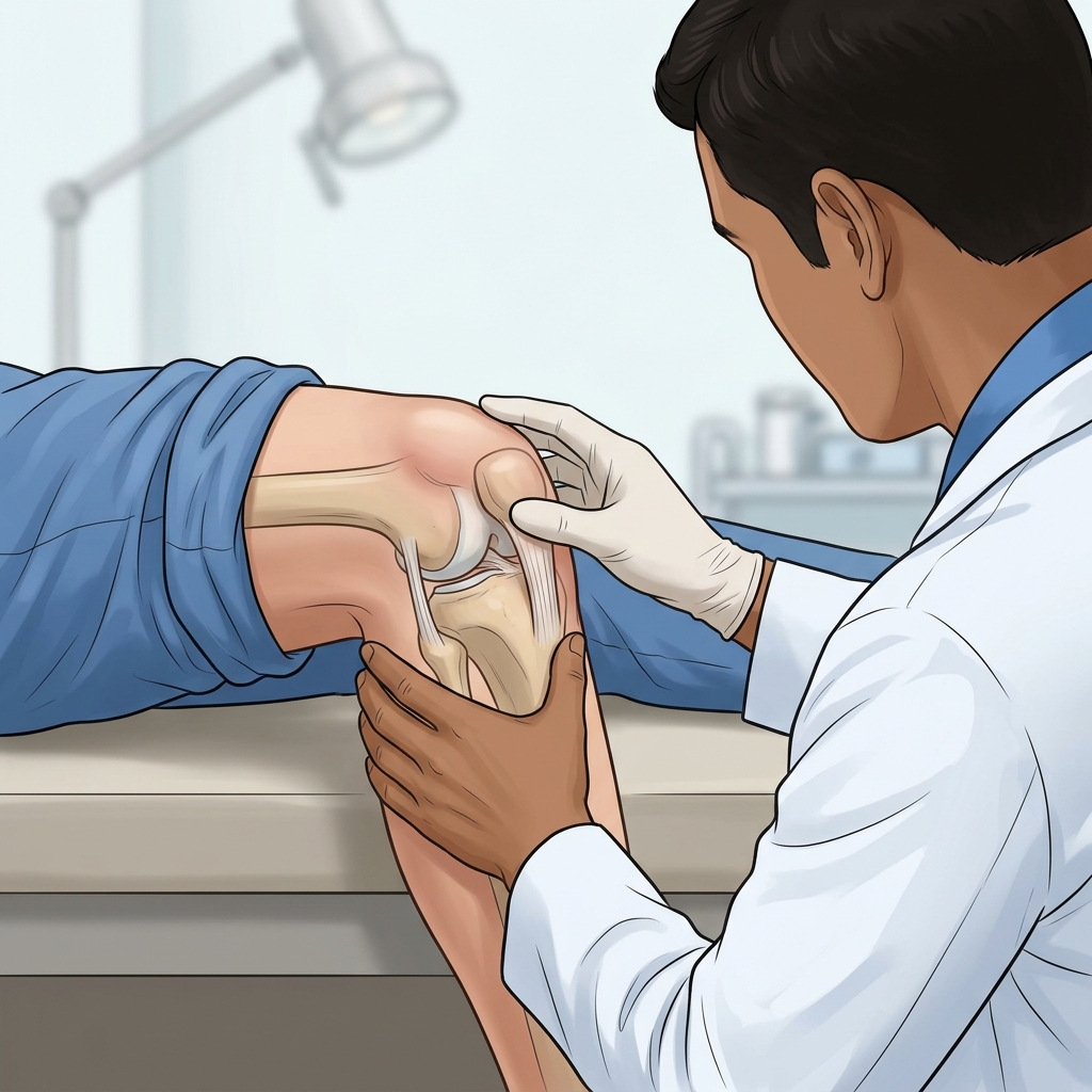
- — Swollen knee arthritis requires both a precise diagnosis and immediate mechanical relief by removing the excess fluid.
- — Differentiating between osteoarthritis wear-and-tear and gout crystal formation is essential for determining the right long-term treatment.
- — Our specialists use advanced MSK ultrasound guidance to guarantee exact needle placement, maximizing fluid extraction while minimizing pain.
- — Joint aspiration provides instant relief from the tightness and pressure that traditional pills simply cannot achieve.
Swollen Knee Arthritis: Diagnosis & Instant Relief
Waking up with a knee that feels stiff, heavy, and filled with fluid can bring your daily routine to a grinding halt. When your knee joint becomes inflamed, the protective synovial membrane overproduces fluid, leading to a condition commonly known as joint effusion. If you are experiencing a severely swollen knee cap, finding out exactly what is going on beneath the surface is critical to your recovery.
Whether the fluid accumulation is due to age-related wear-and-tear or a sudden crystal attack, swollen knee arthritis is a condition that demands immediate attention. Unlike taking oral anti-inflammatory pills—which can take days to work, upset your stomach, and only mask the pain—a therapeutic joint aspiration directly addresses the root of the discomfort by physically removing the trapped fluid and providing instantaneous mechanical relief.
Understanding the Swollen Knee Causes: How to Tell the Difference
Not all swollen knees are created equal. While the physical sensation of tightness might feel similar, the underlying biochemical process causing the fluid buildup varies drastically. The fluid that fills your joint holds the key to your diagnosis. When our specialists evaluate your knee, they are primarily looking to differentiate between three major culprits of sudden joint swelling. Recognizing these symptoms at home can help you understand the urgency of your condition.
1. Degenerative Wear-and-Tear (Osteoarthritis)
Osteoarthritis (OA) is a progressive, chronic condition where the protective cartilage at the ends of your bones gradually wears away over years of use. This bone-on-bone friction causes significant mechanical irritation. In response to this friction, the synovial lining produces excess fluid in a desperate, ultimately flawed attempt to lubricate the joint.
OA-related swelling typically presents with a chronic, dull ache, profound stiffness in the morning that loosens up after 30 minutes of movement, and a feeling of "catching," "clicking," or "grinding" when you walk or climb stairs. Patients on forums like Reddit, YouTube, and Inspire often ask about this specific catching sensation when seeking answers for their knee pain. In OA, the swelling often fluctuates depending on your activity level, getting worse after a long day of standing or walking.
2. Crystal-Induced Inflammation (Gout & Pseudogout)
A swollen knee gout flare-up is entirely different from the mechanical wear of OA. Gout occurs when high levels of uric acid in the bloodstream crystallize and deposit directly into the joint space. These microscopic, needle-like crystals trigger a massive, aggressive, and highly localized immune response.
A gout flare-up is usually incredibly sudden. Patients often go to bed feeling completely fine and wake up in the middle of the night with a knee that is intensely painful, visibly red, hot to the touch, and so sensitive that even the weight of a bedsheet is agonizing. Differentiating between a standard OA flare-up and an acute gout attack is critical because the long-term medication and dietary management strategies are vastly different.
3. Septic Arthritis (Medical Emergency)
Though less common, septic arthritis occurs when a bacterial infection enters the joint space. This is a severe medical emergency. The knee will be extremely swollen, intensely red, and radiating heat. Crucially, septic arthritis is almost always accompanied by systemic symptoms such as a high fever, chills, and profound fatigue. If you suspect your swollen knee is infected, immediate medical evaluation and joint aspiration are required to prevent permanent, irreversible destruction of the cartilage within a matter of days.
The Diagnostic Power of Synovial Fluid Analysis
When you present with a painfully swollen knee, our first priority is to alleviate the pressure safely and quickly while securing an accurate diagnosis. A joint aspiration, medically known as arthrocentesis, accomplishes both of these goals simultaneously. By extracting a sample of the synovial fluid, our specialists can visually and microscopically analyze it. In my clinic, I look for specific signs within the fluid; its color, clarity, and cellular composition tell a detailed story about your joint's internal environment.
| Fluid Characteristic (Visual & Microscopic) | Clinical Indication | Next Steps & Management |
|---|---|---|
| Clear to Pale Yellow, Transparent, Highly Viscous | Normal Joint Fluid or Mild Osteoarthritis | Rest, physical therapy, conservative pain management. |
| Deep Yellow, Slightly Cloudy, Reduced Viscosity | Moderate to Severe Osteoarthritis Flare-Up | Therapeutic steroid injection to calm the synovial lining, followed by viscosupplementation gel lubrication once the joint is stabilized. |
| Opaque, Milky, or Chalky White with Visible Crystals | Gout or Pseudogout Attack | Crystal analysis, localized steroid injection, long-term uric acid management. |
| Bloody (Hemarthrosis), Dark Red or Brown | Acute Trauma (Ligament Tear, Meniscus Tear, Fracture) | Immediate diagnostic MSK ultrasound to assess structural damage, and offloading with custom knee bracing. |
| Thick, Purulent (Pus-like), Yellow/Green, Foul Odor | Septic Arthritis (Bacterial Infection) | Immediate antibiotic therapy, lab cultures, possible surgical washout. |
The Procedure: Step-by-Step Ultrasound-Guided Aspiration
Patients often describe the sensation of a swollen knee as an unbearable tightness, stating they can "barely walk or put weight on the knee." This is because the trapped fluid is exerting immense physical pressure on the surrounding tissues, ligaments, and nerves. Draining the fluid provides profound relief that cannot be matched by ice packs or pain relievers.
In many traditional clinical settings, doctors perform joint aspirations "blind," relying entirely on touch and anatomical landmarks to guess where the fluid pocket is located. This outdated approach can lead to multiple painful needle pricks, accidental damage to the cartilage, and a failure to extract all of the problematic fluid, especially if the fluid is loculated (trapped in small compartments).
At our clinic, we take a superior, modern approach. By utilizing advanced, high-resolution MSK (Musculoskeletal) Ultrasound, we visualize the internal structures of your knee in real-time. Here is exactly what you can expect during the procedure:
- Diagnostic Scanning: Before any needles are prepared, we use the ultrasound probe to scan the entire knee. We locate the exact pockets of fluid, measure the depth, and assess the surrounding cartilage and tendons for any structural damage.
- Sterile Preparation: The skin over the knee is thoroughly cleaned with medical-grade antiseptic solutions (like chlorhexidine or iodine) to create a completely sterile field, eliminating the risk of introducing bacteria into the joint.
- Local Anesthesia: We inject a small amount of local anesthetic (lidocaine) into the skin and the tissue track leading down to the joint capsule. This ensures that the actual aspiration needle causes minimal discomfort.
- Precision Needle Placement: Watching the ultrasound screen in real-time, the specialist guides the aspiration needle directly into the largest pocket of fluid. We can see the needle tip with sub-millimeter precision, ensuring it never scrapes against your sensitive bone or cartilage.
- Complete Fluid Extraction: We gently pull back on the syringe to extract the fluid. Because we can see the fluid pockets shrinking on the ultrasound, we can gently maneuver the needle or apply external pressure to the knee to ensure we evacuate as much fluid as possible. It is not uncommon to extract anywhere from 15 mL to 60 mL of fluid from a severely swollen knee.
- Targeted Therapeutic Injection (Optional but Recommended): Once the fluid is drained, we often leave the needle in place and swap the syringe to inject a precise dose of a corticosteroid (usually 20 mg to 40 mg) directly into the now-empty joint space. This powerfully suppresses the inflamed synovial membrane, preventing the fluid from rapidly re-accumulating the next day.
Post-Procedure Care and Long-Term Rehabilitation
The relief following a successful joint aspiration is often immediate. Patients who limped into the clinic are frequently able to walk out with a significantly improved gait and dramatically reduced pain. However, the aspiration is only the first step in your recovery journey. Proper post-procedure care is vital to ensure the inflammation stays away.
The First 48 Hours
Immediately following the procedure, the knee will be bandaged. We recommend that patients take it easy for the first 24 to 48 hours. While you can walk on the leg, you should strictly avoid strenuous activities, deep squats, heavy lifting, or prolonged periods of standing. Applying an ice pack to the knee for 15 minutes every few hours can help reduce any residual inflammation from the needle track. If a steroid was injected, it may take 24 to 72 hours for the medication to reach its peak anti-inflammatory effect.
Physical Therapy and Strengthening
If your swelling was caused by osteoarthritis, the underlying mechanical instability of the joint remains even after the fluid is gone. To prevent future flare-ups, you must improve the biomechanics of your knee. This is where targeted physical therapy and quad strengthening becomes essential.
A structured rehabilitation program focuses on strengthening the quadriceps (the muscles on the front of your thigh) and the hamstrings (the muscles on the back). Stronger leg muscles act as dynamic shock absorbers, taking the physical load off the damaged cartilage and reducing the bone-on-bone friction that triggers the synovial lining to produce excess fluid. Additionally, improving joint proprioception (your body's awareness of its position in space) and flexibility can significantly extend the lifespan of your natural knee joint.
Transparent Pricing & PhilHealth Coverage
We believe in complete financial transparency for our patients in Vigan City. A standalone ultrasound-guided joint aspiration is ₱2,999. If a therapeutic steroid injection is required after drainage to stop severe inflammation, the total procedure cost is ₱5,999. Both of these procedures are fully covered by PhilHealth for eligible members, meaning you can receive expert care with zero out-of-pocket costs.
Don't let fluid build up and permanently damage your cartilage. The longer toxic enzymes from inflammation remain trapped in your joint, the more they aggressively break down healthy tissue. Located in Vigan, Ilocos Sur, our clinic is fully equipped to handle joint effusions safely and immediately. Accurate diagnosis and swift, ultrasound-guided therapeutic drainage are the cornerstones of protecting your joint health on and beyond. Book your diagnostic MSK ultrasound consultation today to find out exactly what's causing your pain and get the instant relief you deserve.
References & Clinical Evidence
- [1] Sukerkar PA, Doyle Z. Imaging of Osteoarthritis of the Knee. Radiol Clin North Am. 2022 Jul;60(4):583-596. doi: 10.1016/j.rcl.2022.03.004. PMID: 35672093
- [2] Dainese P, Wyngaert KV, De Mits S, Wittoek R, Van Ginckel A, Calders P. Association between knee inflammation and knee pain in patients with knee osteoarthritis: a systematic review. Osteoarthritis Cartilage. 2022 Apr;30(4):516-534. doi: 10.1016/j.joca.2021.12.003. PMID: 34968719
- [3] Gupta PK, Maheshwari S, Cherian JJ, et al. Efficacy and Safety of Stempeucel in Osteoarthritis of the Knee: A Phase 3 Randomized, Double-Blind, Multicenter, Placebo-Controlled Study. Am J Sports Med. 2023;51(9):2289-2299. doi:10.1177/03635465231180323. PMID: 37366164
* Clinical references are provided to support the medical claims made in this article. TeraCare adheres to evidence-based practices in physical medicine and rehabilitation.

Dr. Ben Rabara
Dr. Ben Rabara is a Board-Certified Physiatrist specializing in Physical Medicine and Rehabilitation. He focuses on non-surgical, precision treatments for musculoskeletal conditions, utilizing advanced diagnostics like MSK Ultrasound.
Medical Disclaimer: The information provided in this article is for educational purposes only and does not substitute for professional medical advice, diagnosis, or treatment. Always consult a qualified physician for your specific health conditions.