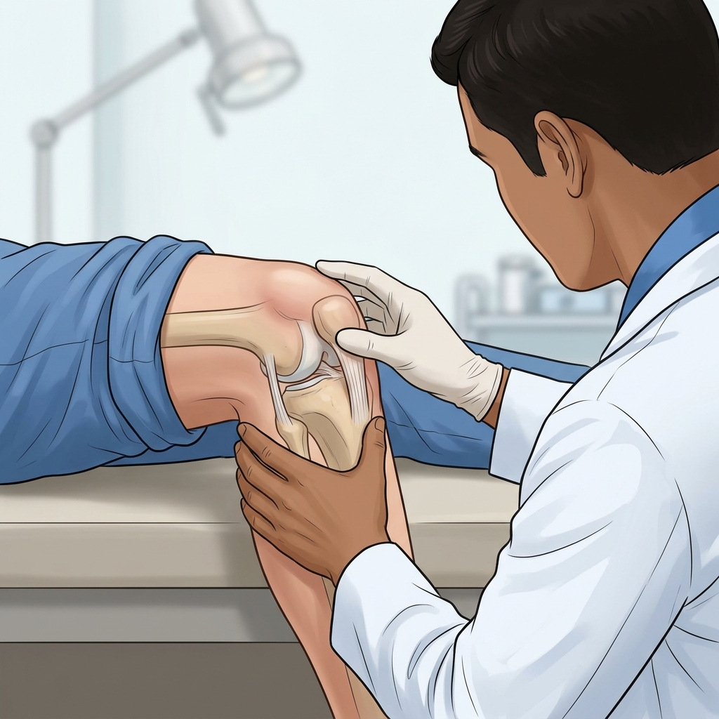
- — A swollen knee (joint effusion) is a sign of underlying joint distress, not merely a symptom to mask with ice.
- — Instead of generic rest, we use dynamic MSK Ultrasound to identify if the fluid is caused by osteoarthritis, a meniscal tear, or inflammation.
- — Pain-free isometric holds can safely stimulate joint health without exacerbating swelling.
- — Targeted, ultrasound-guided therapeutic joint aspirations can safely relieve pressure and pain when conservative care plateaus.
Waking up with a knee that feels like it’s filled with jello—or experiencing a sudden, terrifying ballooning of the joint after an activity—is a jarring experience. Patients often describe the sensation of "water on the knee" as feeling like they knelt in something wet, or they notice the fluid "swishing back and forth" when they try to walk.
Medically known as joint effusion, this localized swelling, or pamamaga, occurs when excess synovial fluid accumulates in or around the knee joint. It is a sign that the joint is in distress. Whether it is a subtle, chronic tightness that limits your ability to climb stairs, or an acute, painful swelling accompanied by a "clicking" or "snapping" sound, understanding why your knee is swollen is the critical first step toward lasting relief.
What Causes Water on the Knee?
The knee joint is encapsulated by a synovial membrane that naturally produces a small amount of fluid to lubricate the cartilage and reduce friction. When the joint is irritated, injured, or diseased, this membrane goes into overdrive, producing excess fluid in an attempt to protect the joint. This protective mechanism, however, leads to the uncomfortable and often painful swelling known as water on the knee.
1. Osteoarthritis and Degenerative Joint Disease
One of the most common causes of chronic knee effusion in older adults is osteoarthritis (often referred to locally as rayuma). As the protective cartilage at the ends of the bones wears down over time, the resulting bone-on-bone friction causes significant irritation. The synovial membrane responds by overproducing fluid. This type of swelling is typically characterized by a chronic, dull ache and significant stiffness, particularly after periods of rest or first thing in the morning.
2. Acute Traumatic Injuries
A sudden twisting motion, a direct blow to the knee, or a hyperextension injury can cause immediate and severe swelling. Common traumatic causes include:
- Ligament Tears: Injuries to the Anterior Cruciate Ligament (ACL) or Medial Collateral Ligament (MCL) often result in rapid swelling (hemarthrosis, or blood in the joint) within hours of the injury.
- Meniscal Tears: The menisci are C-shaped pieces of cartilage that act as shock absorbers between your femur and tibia. A tear can cause localized swelling, often accompanied by a "catching," "clicking," or locking sensation. Patients frequently report a sharp, C-shaped pain on the side of the knee.
3. Overuse Injuries and Bursitis
Repetitive stress on the knee joint can inflame the bursae—small, fluid-filled sacs that cushion the outside of the joint. Prepatellar bursitis (often called "housemaid's knee") causes localized swelling directly over the kneecap. This is common in individuals whose occupations or hobbies require frequent kneeling, and patients often describe it as feeling like they have a pocket of water right on the front of their knee.
4. Inflammatory and Systemic Conditions
Conditions like gout, pseudogout, and rheumatoid arthritis can trigger sudden, severe episodes of joint effusion. These systemic conditions cause the body's immune system to mistakenly attack the joint lining, leading to rapid accumulation of fluid, intense pain, redness, and warmth.
Symptoms of Knee Effusion: Beyond Just Swelling
While the visible enlargement of the knee is the most obvious sign, water on the knee rarely presents in isolation. The specific symptoms accompanying the swelling provide vital clues to the underlying cause:
- Stiffness and Loss of Range of Motion: The excess fluid increases intra-articular pressure, physically blocking the knee from bending or straightening fully.
- Pain and Tenderness: The pain can range from a dull, persistent ache associated with osteoarthritis to sharp, severe pain linked to acute ligament or meniscal tears. The pain may worsen when bearing weight or moving the joint.
- Redness and Warmth: If the swollen knee feels significantly warmer than the surrounding skin or appears red, it may indicate an active inflammatory process (like gout) or, less commonly, an infection.
- Instability or "Giving Way": If the swelling is caused by a structural injury like an ACL tear, the knee may feel wobbly or unstable when you try to stand or walk.
WARNING: If you experience severe pain, an inability to bear weight, a sudden deformity in the knee, or if the swelling is accompanied by a high fever and extreme redness, seek immediate medical attention. These can be signs of a serious structural injury or a dangerous joint infection (septic arthritis).
Diagnostic Precision: The Role of MSK Ultrasound
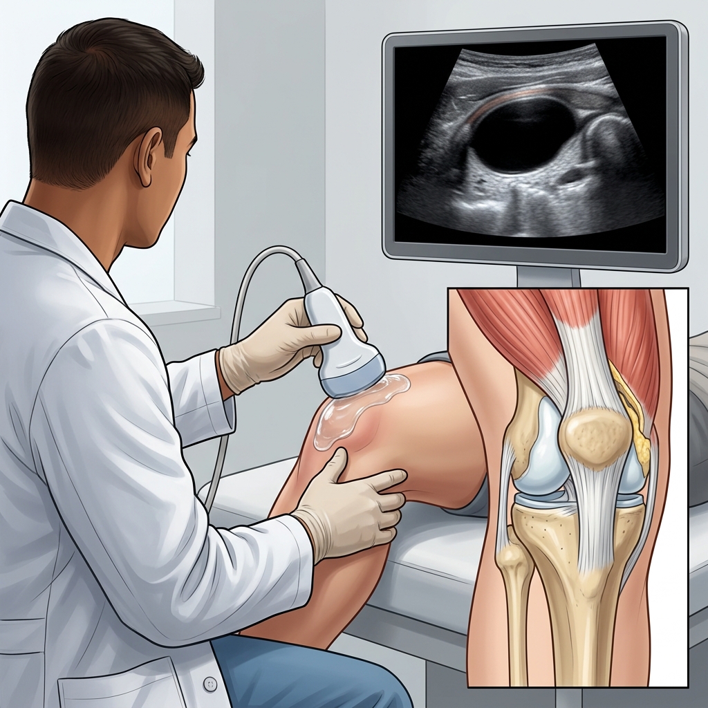
A critical gap in standard clinical assessments is the reliance on static imaging or generic physical exams. At TeraCare Rehabilitation, we utilize dynamic, in-motion Musculoskeletal (MSK) Ultrasound to provide immediate, pinpoint diagnostic accuracy.
While an X-ray is excellent for viewing bones, it cannot visualize soft tissues or fluid. MRI is comprehensive but expensive, time-consuming, and static. MSK Ultrasound bridges this gap perfectly. Under the guidance of a Physical Medicine and Rehabilitation (PM&R) specialist (Physiatrist), ultrasound allows us to:
- Visualize the Fluid in Real-Time: We can directly see the volume and location of the effusion, distinguishing between intra-articular fluid (inside the joint capsule) and extra-articular fluid (like bursitis).
- Assess Soft Tissue Integrity: We can dynamically evaluate the ligaments, tendons (or litid), and menisci while you move your knee, identifying tears, tendinopathy, or impingements that a static image might miss.
- Guide Interventions Safely: If a therapeutic joint aspiration is necessary, ultrasound guidance ensures absolute precision, drastically reducing procedural pain and minimizing the risk of complications.
Research supports this approach. A 2024 meta-analysis in Arthritis Care & Research (PMID: 37522287) confirmed that ultrasound demonstrates promising accuracy in detecting synovial abnormalities, providing equivalent information to MRI but being far more accessible and cost-effective for the patient.
The Treatment Protocol: Moving Beyond R.I.C.E.
The generic advice for a swollen joint has long been the R.I.C.E. protocol: Rest, Ice, Compression, and Elevation. However, modern rehabilitation science has shown that absolute rest is often counterproductive.
The Tactical Home Care Pivot
Complete rest induces rapid muscle atrophy (weakening) and causes the collagen fibers in your tendons to become disorganized. Furthermore, excessive icing causes vasoconstriction, which limits the blood flow required to deliver oxygen and macrophages that help clear debris and synthesize new tissue.
Instead of generic rest, we advocate for active mechanical loading within a safe, pain-free envelope.
- Pain-Free Isometric Holds: By contracting the quadriceps and hamstrings without moving the knee joint (isometrics), you trigger cortical inhibition—a natural analgesic effect. More importantly, this stimulates mechanotransduction, a cellular process that aligns collagen fibers and maintains tendon strength without risking joint shear. Try holding a gentle muscle contraction for 30-45 seconds, 4-5 times a day.
- Light Active Range of Motion: Gently bending and straightening the knee within a range that does not increase pain helps maintain joint lubrication and prevents capsular stiffness.
TIP: Avoid traditional deep tissue massage or local hilot directly over an acutely swollen joint. Aggressive manipulation of a highly inflamed capsule can exacerbate the effusion and increase pain.
Advanced Rehabilitation Modalities
If conservative home management is insufficient, our clinic employs a multimodal approach to reduce swelling and restore function:
- Therapeutic Ultrasound and Laser Therapy: Clinical studies (including a 2025 network meta-analysis, PMID: 40531843) indicate that targeted modalities like therapeutic ultrasound and High-Intensity Laser Therapy (HILT) can effectively reduce inflammation, manage pain, and promote tissue healing when combined with proper exercise.
- Targeted Kinesiology Taping and Bracing: Specialized taping techniques can help improve lymphatic drainage to reduce swelling, while unloader braces can temporarily relieve pressure on damaged cartilage during the healing phase.
Therapeutic Joint Aspirations (Arthrocentesis)
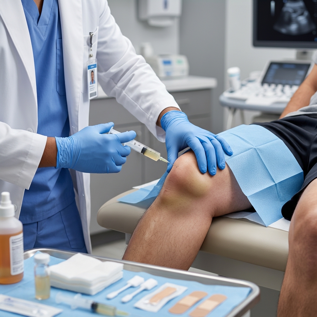
When a knee is severely swollen, tight, and profoundly painful—and conservative measures have plateaued—a therapeutic joint aspiration may be indicated.
Performed by our PM&R specialist under continuous ultrasound guidance, this procedure involves carefully inserting a fine needle into the joint capsule to aspirate (drain) the excess fluid.
The benefits are twofold:
- Immediate Relief: Removing the fluid instantly reduces intra-articular pressure, immediately alleviating the "tight" sensation and restoring a significant degree of mobility.
- Diagnostic Value: The aspirated fluid can be analyzed to determine the exact cause of the swelling (e.g., checking for uric acid crystals in gout, or signs of infection).
In some cases, after the fluid is drained, a corticosteroid or hyaluronic acid injection may be administered directly into the joint to suppress further inflammation and provide longer-lasting relief. Recent guidelines (PMID: 40015722) emphasize that ultrasound guidance significantly increases the accuracy of these injections and reduces procedural discomfort.
Navigating Care in Northern Luzon
We understand that seeking specialized care in the provinces presents unique challenges. Traveling from remote municipalities to Vigan City or Bantay while dealing with a painful, swollen knee can be daunting.
At TeraCare Rehabilitation, we prioritize diagnostic efficiency. By utilizing in-clinic MSK Ultrasound, we often eliminate the need for patients to travel to larger hospitals for expensive MRI scans. This "one-stop" diagnostic and treatment approach saves you time, reduces travel burden, and lowers overall out-of-pocket costs. Furthermore, we maintain transparent pricing models and can assist in navigating PhilHealth coverage options for eligible outpatient procedures to ensure you receive the high-quality care you deserve without unnecessary financial stress.
Do not let a swollen knee dictate your mobility. With the right diagnostic precision and a targeted rehabilitation protocol, you can extinguish the pain and return to the activities you love.
Get Relief from Joint Swelling
Schedule an ultrasound-guided joint evaluation with Dr. Rabara.
References & Clinical Evidence
- [1] Liu K, et al. Diagnostic Accuracy of Ultrasound for Assessment of Synovial Abnormalities Among Patients With Knee Pain: A Meta-Analysis. Arthritis Care Res (Hoboken). 2024. PMID: 37522287.
- [2] Aiyer R, et al. Therapeutic Ultrasound for Chronic Pain Management in Joints: A Systematic Review. Pain Med. 2020. PMID: 31095336.
- [3] Chen X, et al. Clinical efficacy of different therapeutic options for knee osteoarthritis: A network meta-analysis based on randomized clinical trials. PLoS One. 2025. PMID: 40531843.
- [4] Benzon HT, et al. Use and safety of corticosteroid injections in joints and musculoskeletal soft tissue: guidelines from the American Society of Regional Anesthesia and Pain Medicine. Reg Anesth Pain Med. 2025. PMID: 40015722.
* Clinical references are provided to support the medical claims made in this article. TeraCare adheres to evidence-based practices in physical medicine and rehabilitation.

Dr. Ben Rabara
Dr. Ben Rabara is a Board-Certified Physiatrist specializing in Physical Medicine and Rehabilitation. He focuses on non-surgical, precision treatments for musculoskeletal conditions, utilizing advanced diagnostics like MSK Ultrasound.
Medical Disclaimer: The information provided in this article is for educational purposes only and does not substitute for professional medical advice, diagnosis, or treatment. Always consult a qualified physician for your specific health conditions.