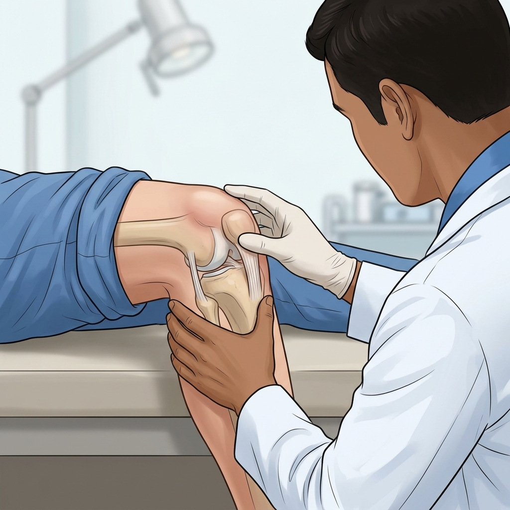
- — Home remedies like Rest, Ice, and Elevation provide temporary superficial pain relief, but cannot physically evacuate high-pressure fluid trapped inside a closed joint capsule.
- — Applying tight elastic compression wraps over a tense joint effusion can paradoxically increase intra-articular pressure, worsening quadriceps muscle inhibition.
- — Intra-articular pressure above 50 mmHg triggers Arthrogenic Muscle Inhibition (AMI), shutting down quadriceps firing by up to 90% until the pressure is mechanically relieved.
- — Ultrasound-guided joint aspiration (arthrocentesis) acts as an immediate pressure-release valve, dropping internal joint pressure back to normal sub-atmospheric levels in minutes.
Waking up to find your knee enlarged, tight, and resistant to bending can turn even basic daily activities into an agonizing ordeal. Scientifically termed joint effusion—and colloquially referred to as "water on the knee"—this condition occurs when the synovial membrane lining your joint capsule experiences severe inflammation and overproduces fluid. If you are frantically searching for how to treat water on the knee or wondering whether your knee swelling home remedy will ever work, understanding the biomechanics of fluid trapped inside a sealed joint capsule is the essential first step toward lasting recovery.
Many patients spend weeks resting, elevated, and wrapped in elastic bandages, hoping the fluid will simply absorb back into their body. In my clinic, I look for signs of severe capsular pressure when patients arrive complaining of a knee that feels stiff, heavy, or locked. On patient community forums like Reddit and YouTube, people frequently describe this as feeling like "a water balloon stuck inside the joint" that produces a dull, burning ache or a weak, catching sensation when walking. While superficial conservative measures have an important initial role in acute symptom control, high-volume joint effusions create a physical pressure barrier that home remedies cannot cross. Below, we examine the physics of intra-articular pressure, evaluate traditional home remedies against medical fluid drainage (arthrocentesis), and outline when immediate clinical intervention is required to protect your joint cartilage.
Ready for Immediate Pressure Relief?
Don't let high-pressure fluid destroy your knee cartilage. Schedule your diagnostic MSK ultrasound consultation with Dr. Rabara today.
The Physics of "Water on the Knee": Intra-Articular Pressure & Joint Capsule Mechanics
When I evaluate a patient presenting with a severely swollen knee, the first thing I explain is the anatomy of the knee joint capsule. The knee is an airtight, sealed synovial chamber designed to hold approximately 2 to 5 mL of clear lubricating fluid under normal conditions. In a healthy joint, intra-articular pressure (IAP) remains slightly sub-atmospheric (ranging from -2 to +2 mmHg during rest), allowing the joint surfaces to glide effortlessly without capsular tension.
When joint structures suffer mechanical trauma (such as a meniscus tear or cartilage breakdown) or biochemical irritation (such as a gout flare-up), the synovial lining shifts into hyper-secretion mode. As fluid volume escalates from 5 mL up to 30, 60, or even 100 mL, the fibrous joint capsule stretches to its absolute elastic limit. This dramatic volume expansion causes intra-articular pressure to spike exponentially, frequently exceeding 50 mmHg.
High intra-articular pressure triggers a destructive physiological cascade known as Arthrogenic Muscle Inhibition (AMI). As capsular tension rises, specialized mechanoreceptors embedded within the joint capsule fire continuous high-frequency inhibitory signals directly to the spinal cord. These signals activate spinal Ib non-reciprocal inhibitory interneurons, which selectively suppress the motor neuron pool governing the quadriceps muscle. Clinical studies demonstrate that tense knee effusions can shut down quadriceps voluntary muscle activation by 30% to 90%, leading to rapid muscle wasting, severe gait limping, and a feeling of weak, un-steady knees with tingling or sharp radiating discomfort.
The RICE Protocol: What Works, What Fails, and the Paradoxical Risk of Compression
In my rehabilitation practice, I am constantly asked about the RICE protocol (Rest, Ice, Compression, Elevation). While it is universally recommended as a standard first-aid response, I always caution my patients that when applied to a high-volume, high-pressure intra-articular knee effusion, each component of RICE has strict physiological limitations that you must understand.
1. Rest (R)
What works: Halting high-impact activities, heavy lifting, and deep squatting prevents further mechanical tearing of damaged cartilage or meniscal tissue, stopping the initial surge of inflammatory signaling.
What fails: Passive rest does not actively reduce fluid volume or lower intra-articular pressure. Furthermore, prolonged immobility in the setting of persistent joint effusion accelerates quadriceps muscle atrophy and joint stiffness without facilitating fluid reabsorption.
2. Ice / Cryotherapy (I)
What works: Applying cold packs for 15 to 20 minutes every 2 to 4 hours causes cutaneous vasoconstriction, slows local nerve conduction velocity, and decreases inflammatory metabolic demand, providing noticeable superficial pain relief.
What fails: Cold temperatures affect only the superficial skin and subcutaneous tissues. Ice cannot penetrate deep inside the sealed joint capsule to alter fluid volume or reduce internal capsular pressure.
3. Compression (C) — The Paradoxical Risk
What works: Light compression wrap on a low-grade extra-articular strain can reduce subcutaneous tissue edema by favoring lymphatic uptake.
What fails & The Paradoxical Risk: Applying a tight elastic compression wrap over a severely swollen, high-volume intra-articular effusion is clinically counterproductive. Because the joint capsule is already stretched to maximum capacity, applying external compressive force pushes against a non-compressible fluid column. This paradoxically increases intra-articular pressure further, exacerbating mechanoreceptor firing, worsening Arthrogenic Muscle Inhibition, and driving severe ischemic pain. In my clinic, I explicitly advise patients to remove tight elastic wraps if their knee feels increasingly tight or painful.
4. Elevation (E)
What works: Elevating the affected leg above heart level utilizes gravity to assist venous return and lymphatic drainage from the lower leg, ankle, and foot.
What fails: While elevation helps clear interstitial fluid in soft tissues, the synovial capsule is a distinct anatomical compartment isolated from general interstitial space. Highly inflamed, congested capsular lymphatic channels cannot drain large volumes of intra-capsular fluid through passive gravity alone.
The Over-Inflated Tire Analogy: Why Surface Remedies Cannot Shrink High-Pressure Effusions
Consider a car tire that has been over-inflated to double its maximum rated pressure. If you attempt to reduce the internal air pressure by applying ice packs to the rubber tread, wrapping duct tape tightly around the outside of the tire, or letting the car sit parked in the garage, the internal air pressure remains completely unchanged. The only way to lower the pressure inside an over-inflated tire is to depress the valve stem and allow the air to escape mechanically.
A high-volume swollen knee operates under the exact same fluid dynamics. The joint capsule is the sealed tire, and the excess synovial fluid is the over-pressurized air. Ice, rest, and elevation act only on the outer rubber (the skin and superficial tissues). When internal joint pressure exceeds 50 mmHg, surface remedies reach a physiological ceiling. Until a physician depresses the "valve stem"—performing an ultrasound-guided joint aspiration—the trapped fluid will continue to stretch the joint capsule, destroy healthy cartilage through exposure to toxic inflammatory cytokines, and paralyze your quadriceps.
Knee Fluid Aspiration (Arthrocentesis): The Mechanical Pressure Valve
Knee joint aspiration, known medically as arthrocentesis, is a minor outpatient procedure where I use a sterile needle under real-time ultrasound visualization to evacuate excess synovial fluid directly from your joint space. Rather than forcing you to wait weeks for passive, incomplete lymphatic reabsorption, aspiration acts as an immediate mechanical pressure-release valve.
The physiological transformation following joint fluid evacuation is dramatic and immediate:
- Instant Pressure Drop: Intra-articular pressure drops from toxic levels (above 50 mmHg) back to normal sub-atmospheric resting baseline (0 to 2 mmHg) within seconds of fluid withdrawal.
- Immediate Quads Unlocking: Neutralizing capsular tension turns off the inhibitory spinal reflex (AMI), immediately re-establishing quadriceps motor unit recruitment and restoring leg strength.
- Mechanical Range of Motion Restoration: Removing 30 to 60 mL of trapped fluid removes the physical barrier to joint flexion, allowing patients to bend and extend their knee without feeling a tight "bursting" sensation.
- Cytokine Washout: Synovial fluid in a swollen knee contains high concentrations of destructive catabolic enzymes (such as Matrix Metalloproteinases MMP-1 and MMP-13) and pro-inflammatory cytokines (IL-1β, TNF-α). Extracting this toxic fluid physically clears these destructive agents out of the joint, protecting remaining articular cartilage from rapid enzymic breakdown.
Comparative Matrix: Home Remedies vs. Ultrasound-Guided Aspiration
The table below provides a clear, clinical comparison evaluating traditional home self-care against professional ultrasound-guided joint aspiration across key therapeutic dimensions:
| Clinical Feature / Metric | Home Remedies (RICE & OTC Pills) | Ultrasound-Guided Aspiration (Arthrocentesis) |
|---|---|---|
| Primary Mechanism | Superficial analgesia & mild cutaneous vasoconstriction | Direct mechanical fluid removal & intra-articular pressure collapse |
| Intra-Articular Pressure Impact | Zero reduction in capsular hydrostatic pressure | Instant drop from above 50 mmHg to sub-atmospheric (0 to 2 mmHg) |
| Speed of Pressure Relief | Slow (days to weeks, often incomplete) | Immediate (occurs within minutes during procedure) |
| Impact on Quads Inhibition (AMI) | No effect; quadriceps remain inhibited by capsular stretch | Abolishes AMI reflex, immediately restoring quadriceps motor firing |
| Diagnostic Capability | None; cannot identify underlying fluid composition | High diagnostic yield (synovial fluid crystal, cell count & culture analysis) |
| Cartilage Protection | Low; toxic cytokines remain trapped against cartilage | High; physically evacuates inflammatory enzymes and blood breakdown products |
| Procedure Guidance & Safety | N/A (Self-administered) | Sub-millimeter real-time MSK Ultrasound needle visualization (POCUS) |
Conditions Commonly Mistaken for Knee Effusion
When patients consult me for knee swelling, my immediate diagnostic priority is confirming whether the fluid is actually located *inside* the joint capsule. On discussion forums such as Reddit and Inspire, patients often confuse true joint swelling with other soft tissue conditions that present with visible knee enlargement but require completely different treatment strategies:
1. Prepatellar Bursitis ("Housemaid's Knee")
Prepatellar bursitis is fluid accumulation inside the superficial bursa located directly in front of the kneecap. It appears as a localized, dome-shaped "egg" sitting on top of the patella. As explained in our guide to tendonitis vs bursitis ultrasound diagnosis, prepatellar bursitis is extra-articular; the knee joint itself is not pressurized, patellar tapping is negative, and patients can often achieve full knee flexion without deep joint tightness.
2. Baker's Cyst (Popliteal Cyst)
A Baker's cyst is a posterior herniation of the joint capsule into the popliteal fossa behind the knee. It occurs when chronic intra-articular pressure forces synovial fluid through a one-way valve between the semimembranosus and gastrocnemius bursae. Patients feel extreme tightness in the back of the knee and upper calf. Treating a Baker's cyst directly by popping or aspirating the posterior cyst alone usually fails; the primary intra-articular joint effusion must be drained from the front to collapse the posterior valve.
3. Pes Anserine Bursitis
Pes anserine bursitis is an inflammation of the bursa located on the inner, lower aspect of the knee (approximately 2 inches below the medial joint line). It causes localized point tenderness over the upper tibia, particularly in runners or patients with knock-knee alignment, but does not cause floating kneecaps or generalized intra-articular pressure.
Clinical Evidence & Common Misconceptions
In my clinic, I look for opportunities to educate patients on modern evidence-based physiatry, which challenges several widespread clinical myths regarding swollen knee management:
Myth 1: "Draining a knee causes the body to produce even more fluid."
Clinical Reality: Aspiration does not stimulate fluid production. The synovial membrane overproduces fluid because of an unaddressed underlying trigger (such as active gout crystals, a torn meniscus, or degenerative cartilage degradation). If the underlying cause remains unmanaged, fluid will naturally re-accumulate over time. This is why our physiatrists combine fluid aspiration with real-time diagnostic ultrasound scanning and, when appropriate, a targeted anti-inflammatory corticosteroid injection to shut down synovial hyper-secretion at its source. For chronic bone-on-bone friction, we subsequently transition patients to long-term lubrication via viscosupplementation gel injections.
Myth 2: "Physical examination alone is sufficient to diagnose knee fluid."
Clinical Reality: Clinical physical exam maneuvers (such as the patellar tap test or bulge sign) have surprisingly poor diagnostic specificity (31% to 37%) and miss subtle or loculated fluid collections. Point-of-Care Musculoskeletal Ultrasound (POCUS) detects joint effusions down to 1 to 2 mL with over 95% diagnostic accuracy, preventing unnecessary needle insertions when fluid is absent or extra-articular.
Transparent Pricing & PhilHealth Coverage
We maintain absolute financial transparency for all medical diagnostic and therapeutic procedures at our clinic in Vigan City:
- Standalone Ultrasound-Guided Joint Aspiration: ₱2,999 (Includes real-time POCUS diagnostic scanning and complete fluid evacuation).
- Combined Aspiration + Therapeutic Anti-Inflammatory Injection: ₱5,999 (Includes joint decompression followed by targeted corticosteroid administration to stop fluid re-accumulation).
- PhilHealth Coverage: Both procedures are 100% fully covered by PhilHealth for eligible members presenting with medical indications, resulting in zero out-of-pocket procedure costs.
Schedule Your Joint Fluid Evaluation
Accurate diagnosis and swift, ultrasound-guided therapeutic drainage are the cornerstones of protecting your joint health.
Clinical Realities of Knee Fluid Management
Understanding what home remedies can and cannot achieve for joint effusion.
Lymphatic Drainage Limitations
The lymphatic vessels surrounding an inflamed joint capsule quickly become congested. Once fluid accumulation exceeds 15–20 mL, passive lymphatic reabsorption slows dramatically.
Arthrogenic Muscle Inhibition (AMI)
Trapped high-pressure fluid acts as a neural kill-switch for your quadriceps. Exercising through a swollen joint without draining it accelerates muscular atrophy.
Underlying Pathology Persistence
Draining the fluid relieves the immediate mechanical pressure, but long-term success requires diagnosing and treating the primary trigger—whether degenerate cartilage, a torn meniscus, or uric acid crystals.
References & Clinical Evidence
- [1] Elsawy AM, et al. Diagnostic accuracy of point-of-care ultrasound versus clinical physical examination for detection of knee joint effusion. BMC Musculoskelet Disord. 2023. PMID: 10257372.
- [2] Dainese P, Wyngaert KV, De Mits S, Wittoek R, Van Ginckel A, Calders P. Association between knee inflammation and knee pain in patients with knee osteoarthritis: a systematic review. Osteoarthritis Cartilage. 2022 Apr;30(4):516-534. doi: 10.1016/j.joca.2021.12.003. PMID: 34968719.
- [3] Sukerkar PA, Doyle Z. Imaging of Osteoarthritis of the Knee. Radiol Clin North Am. 2022 Jul;60(4):583-596. doi: 10.1016/j.rcl.2022.03.004. PMID: 35672093.
- [4] Gupta PK, Maheshwari S, Cherian JJ, et al. Efficacy and Safety of Stempeucel in Osteoarthritis of the Knee: A Phase 3 Randomized, Double-Blind, Multicenter, Placebo-Controlled Study. Am J Sports Med. 2023;51(9):2289-2299. PMID: 37366164.
* Clinical references are provided to support the medical claims made in this article. TeraCare adheres to evidence-based practices in physical medicine and rehabilitation.

Dr. Ben Rabara
Dr. Ben Rabara is a Board-Certified Physiatrist specializing in Physical Medicine and Rehabilitation. He focuses on non-surgical, precision treatments for musculoskeletal conditions, utilizing advanced diagnostics like MSK Ultrasound.
Medical Disclaimer: The information provided in this article is for educational purposes only and does not substitute for professional medical advice, diagnosis, or treatment. Always consult a qualified physician for your specific health conditions.