- — Corticosteroid injections function like joint fire extinguishers; they suppress severe pain temporarily but are chondrotoxic and accelerate cartilage degeneration if repeated.
- — Arthroscopic meniscus surgeries or 'cleanups' for degenerative tears increase peak contact pressures on cartilage by up to 200%, accelerating the onset of bone-on-bone osteoarthritis.
- — High-concentration autologous Platelet-Rich Plasma (PRP) acts as a biological modifier, delivering essential growth factors (PDGF, TGF-β, VEGF) under live ultrasound guidance to shift joint chemistry toward healing and avoid replacement.
- — Successful joint preservation requires pairing precise biological injections with progressive mechanical loading—a structured rehabilitation program to guide new tissue remodeling.
Many patients in the Philippines diagnosed with rayuma sa tuhod (knee osteoarthritis) or a torn meniscus are quickly guided toward the operating room. The prospect of undergoing a total knee replacement or arthroscopic surgery often triggers deep anxiety, not just about the surgical risks and hospital stays, but also regarding the extensive out-of-pocket costs and the long, painful recovery. Many patients in Vigan and the wider Ilocos region wonder if there is an alternative that allows them to preserve their natural joint structure and avoid surgery entirely.
Platelet-Rich Plasma (PRP) therapy has emerged as a leading non-surgical joint preservation treatment in physical medicine and rehabilitation (physiatry). Rather than merely masking pain or mechanically swapping the joint with metal and plastic, PRP uses the patient’s own biological growth factors to alter the joint environment, stabilize partial meniscus tears, and cool down chronic inflammation. This guide outlines the clinical mechanics, evidence-based outcomes, and practical rehabilitation pathways for patients seeking to avoid knee surgery.
If your main concern is lubricant-style symptom relief or delaying knee replacement with a mechanical cushion, compare PRP with our hyaluronic acid gel alternatives guide.
To understand the real-life struggles of those dealing with joint wear, I regularly review patient discussions and forum comments on online communities like Reddit (such as patient posts on orthopedic recovery subreddits), YouTube comment sections, and health forums (such as Inspire and PatientsLikeMe). In my clinic in Vigan, I recommend an individualized assessment to understand what patients actually share about their daily limitations. Patients often describe knee osteoarthritis pain as a dull, aching sensation deep in the joint that becomes sharp during movement. Degenerative meniscus tears can cause a catching or grinding feeling when bending, accompanied by a stiff, heavy sensation. In some cases, patients also experience a radiating discomfort or a tingling, burning feeling around the joint line when walking, accompanied by a weak feeling in the quadriceps.
What Generic Medical Guides Miss About Knee PRP and Meniscus Tears
In my practice, I find that generic medical websites often oversimplify PRP therapy as a simple injection that magically "cures" joint wear, while ignoring the mechanical forces that caused the tissue breakdown in the first place. These guides fail to explain that biology and mechanics must work together. If a patient receives a PRP injection but continues to walk with poor foot alignment, weak hip stabilizers, or improper knee mechanics, the new healing tissue will be subjected to abnormal shear forces and fail. Biological healing requires mechanical guidance; injection and exercise are not separate options, but partners in joint preservation.
When is Surgery Actually Necessary for Knee Osteoarthritis and Meniscus Tears?
Knee replacement surgery or meniscus repair becomes necessary primarily in end-stage, "bone-on-bone" osteoarthritis (Kellgren-Lawrence Grade 4) with severe mechanical locking, leg collapse, or when conservative treatments fail to restore minimal functional mobility. For mild-to-moderate arthritis (Grades 1-3) and degenerative meniscus tears, non-surgical biological interventions should be exhausted first to preserve native joint tissues. Understanding the specific structural state of your joint is critical to making an informed decision before choosing an invasive procedure.
Clinicians classify joint wear using the Kellgren-Lawrence (KL) grading system, which measures joint space narrowing and bone changes under weight-bearing X-rays:
- Grade 1 (Doubtful): Minute osteophytes (bone spurs) with doubtful joint space narrowing. Surgery is never indicated.
- Grade 2 (Minimal): Clear osteophyte formation and mild narrowing of the joint space. The knee structure is fully intact.
- Grade 3 (Moderate): Moderate joint space narrowing, subchondral sclerosis (thickening of the bone beneath cartilage), and multiple osteophytes. The joint is irritated but retains salvageable cartilage.
- Grade 4 (Severe): Severe joint space narrowing, complete loss of cartilage ("bone-on-bone" contact), large osteophytes, and visible bone deformity. This is the stage where mechanical replacement becomes the primary option.
For patients with Grade 1, 2, or 3 osteoarthritis, or those diagnosed with degenerative meniscus tears, jumping straight to surgery is often premature. In fact, clinical practice guidelines from the European Society of Sports Traumatology, Knee Surgery and Arthroscopy (ESSKA) state that non-surgical conservative treatments should always be the first line of defense for degenerative meniscal tears (Kopf et al., 2020; PMID: 32052121). A degenerative tear is a slow wear-and-tear process associated with aging and joint wear, not a sudden athletic rupture. Treating it surgically by cutting out the tissue often accelerates joint decay rather than resolving the root cause (Ozeki et al., 2022; PMID: 35455094).
The Cortisone Danger: Why Steroids Are a Temporary Fire Extinguisher, Not a Cure
Corticosteroid injections function like joint fire extinguishers, rapidly suppressing severe pain for 4 to 12 weeks, but they do not heal tissues. Repeated steroid injections are clinically associated with chondrotoxicity—accelerating cartilage degeneration and weakening tendons, which ultimately hastens the need for joint replacement. While a steroid shot may offer quick relief during an acute flare-up, it does not support long-term joint health.
Corticosteroids work by shutting down the local inflammatory pathway at a cellular level. However, this suppression comes at a steep biological cost. In vitro and in vivo studies demonstrate that high doses of steroids are toxic to chondrocytes (the cells responsible for producing and maintaining knee cartilage). Repeated injections inhibit these cells from synthesizing collagen and proteoglycans, the building blocks that keep cartilage soft, hydrated, and capable of absorbing shock (Vlad et al., 2024; PMID: 39606697). Over time, this leads to accelerated thinning of the joint space.
Furthermore, steroid injections introduce a dangerous clinical paradox: by turning off the pain alarm without repairing the joint structure, patients often feel a false sense of security. They return to heavy loading, long walks, or physical labor in their farms or businesses while their joint is chemically numb. Because the underlying cartilage damage and meniscus tear are still present, this un-monitored overloading results in micro-trauma, turning partial tears into complete ruptures and driving a Grade 2 knee into a Grade 4 "bone-on-bone" state much faster than normal.
How Platelet-Rich Plasma (PRP) Rebuilds the Joint Environment
Platelet-rich plasma therapy utilizes concentrated growth factors from the patient's own blood to stimulate cellular repair, reduce chronic synovial inflammation, and shift joint biochemistry from catabolism to repair. Under high-resolution ultrasound guidance, PRP is delivered precisely to targeted joint tissues. This autologous approach harnesses your body’s natural healing cascade to stabilize joint environment at a cellular level.
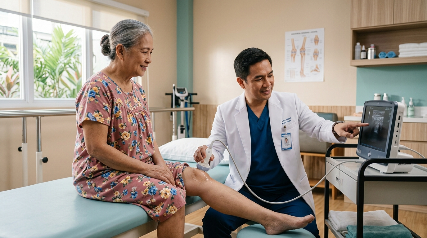
In my assessment of knee pain, I perform a diagnostic musculoskeletal ultrasound to check the tissue state of the joint. Musculoskeletal ultrasound refers to the point-of-care imaging technique that uses sound waves to inspect soft tissues in real-time, as detailed in our guide on namamagang tuhod ultrasound examination. In my practice, I scan the knee joint dynamically to check for a joint effusion. A joint effusion is defined as the abnormal accumulation of fluid inside the joint cavity, which happens due to active inflammation. If the patient has a significant fluid trap, it can be drained via therapeutic joint aspiration (arthrocentesis) under live ultrasound guidance before delivering the platelets.
The clinical preparation of PRP involves drawing approximately 10 to 20 mL of the patient's blood, which is then processed in a specialized medical centrifuge. By spinning the blood at precise gravitational speeds, we separate the red blood cells and plasma, concentrating the platelets up to 5 to 8 times their baseline levels. These concentrated platelets contain alpha-granules that store vital signaling proteins and growth factors, including:
- Platelet-Derived Growth Factor (PDGF): Promotes cell replication, blood vessel formation (angiogenesis), and macrophage activation.
- Transforming Growth Factor-Beta (TGF-β): Stimulates chondrocyte proliferation and collagen synthesis, supporting extracellular matrix health.
- Vascular Endothelial Growth Factor (VEGF): Triggers angiogenesis to bring blood flow and nutrients to areas with poor vascular supply.
- Fibroblast Growth Factor (FGF): Accelerates tissue remodeling and tendon/ligament repair.
When injected into an osteoarthritic knee, PRP does not act by instantly "regrowing" a thick pad of brand-new cartilage. Instead, it acts as a powerful biochemical modulator. The knee joint environment in osteoarthritis is highly catabolic—inflammatory molecules like Interleukin-1 Beta (IL-1β) and Tumor Necrosis Factor-Alpha (TNF-α) dominate, constantly breaking down cartilage matrix. PRP delivers a massive wave of anti-inflammatory cytokines (such as IL-1 Receptor Antagonist) that block these destructive signals, shifting the joint into an anabolic (rebuild and repair) state (Blaga et al., 2024; PMID: 39194659). Synovial fluid inflammation decreases, joint lubrication improves, and the cellular environment stabilizes, allowing patients to experience lasting pain relief and functional restoration (Vlad et al., 2024; PMID: 39606697).
To achieve this, the injection must be delivered with high-precision. "Blind" or landmark-guided injections often misplace the platelets into the surrounding fat pad (Hoffa's fat pad) or joint capsule, causing pain and wasting the biological sample. Using live ultrasound guidance, the physiatrist visualizes the needle in real-time, ensuring the concentrated PRP lands directly in the suprapatellar recess or the joint space adjacent to the damaged meniscus, maximizing clinical efficacy.
Healing Degenerative Meniscus Tears Without Surgery: The Structural Argument
Degenerative meniscus tears do not require surgical removal (partial meniscectomy) in the majority of cases because cutting out meniscus tissue increases pressure on joint cartilage by up to 200%. Autologous PRP injections stabilize the tear site by promoting localized fibrocartilage healing and sealing partial tears, avoiding the accelerated arthritis caused by surgery. Preserving the meniscus is vital to protecting the joint's mechanical lifespan.
The meniscus is a wedge-shaped fibrocartilage pad that acts as a crucial shock absorber, distributing up to 70% of the knee’s load. A meniscus tear is defined as a rupture of this fibrocartilaginous structure due to trauma or wear. When an orthopedic surgeon suggests an arthroscopic "cleanup" or "debridement" for a degenerative tear, they are typically performing a partial meniscectomy—cutting out the torn, frayed portion of the meniscus. While this might sound logical to a patient, it is biomechanically destructive. Removing even 15% to 30% of the meniscus reduces the contact area of the joint, concentrating the patient's entire body weight onto a smaller patch of articular cartilage. This raises peak contact stresses by up to 200%, rapidly grinding down the remaining cartilage and accelerating bone-on-bone friction (Ozeki et al., 2022; PMID: 35455094).
Furthermore, multiple high-quality randomized controlled trials have demonstrated that arthroscopic partial meniscectomy for degenerative meniscus tears offers no long-term benefit over sham surgery (placebo procedures) or structured physical therapy alone. The surgery removes vital mechanical structure without addressing the underlying biochemical wear. PRP therapy takes the opposite approach: it aims to heal and stabilize. The meniscus is divided into zones based on blood supply:
| Zone Name | Vascularity Description | Natural Healing Potential | PRP Treatment Strategy |
|---|---|---|---|
| Red-Red Zone (Outer) | Rich blood supply from joint capsule | High | PRP accelerates and strengthens structural fiber synthesis |
| Red-White Zone (Middle) | Borderline, limited blood flow | Moderate-Low | PRP introduces necessary growth factors to stimulate localized angiogenesis |
| White-White Zone (Inner) | Avascular (no blood supply) | Very Poor | PRP acts as a biochemical modulator to reduce synovial irritation and pain |
By targeting the Red-Red and Red-White border zones under ultrasound guidance, PRP delivers a concentrated dose of growth factors that recruit stem cells, promote angiogenesis, and stimulate fibrocartilage repair (Elphingstone et al., 2024; PMID: 39036807). This biological welding stabilizes the tear, prevents it from propagating, and preserves the shock-absorbing volume of the joint (Li Z et al., 2022; PMID: 36209223).
The Practical Care Pathway: PRP Injections and Rehabilitation in Vigan
Successful non-surgical knee treatment combines a series of 1 to 3 ultrasound-guided PRP injections with a structured, progressive loading physical therapy program. This combined approach ensures that as growth factors stimulate cellular repair, the healing tissues are mechanically stimulated to remodel into strong, functional collagen matrices. Physical rehabilitation is the essential partner to biological healing.
A typical non-surgical joint preservation plan at our Vigan City clinic follows a structured, three-phase care pathway adapted to the physiological stages of tissue repair:
Phase 1: The Inflammatory Phase (Weeks 1-2)
The goal is to protect the joint and allow the concentrated platelets to trigger the initial repair cascade.
- Biological Activity: Platelets release growth factors and initiate a controlled inflammatory response to recruit healing cells.
- Timelines & Rest: Restrict high-impact activities and prolonged standing for the first 48 to 72 hours. Walking around the house is fully allowed, but heavy lifting and running are avoided.
- Friction Alert (No NSAIDs): Patients must avoid taking anti-inflammatory pain relievers (NSAIDs like Ibuprofen, Naproxen, or Mefenamic Acid) for at least 2 weeks after PRP. These drugs block the inflammatory cascade that PRP is trying to initiate. We manage pain using ice and acetaminophen.
- Warning Signs (Red Flags): If you experience high fever, chills, rapidly spreading redness, or severe throbbing pain unresponsive to paracetamol, contact the clinic immediately to rule out infection.
Phase 2: The Proliferation Phase (Weeks 3-6)
The goal is to introduce controlled mechanical loading to guide the alignment of new collagen fibers.
- Biological Activity: Fibroblasts lay down new collagen type I and III fibers, creating new tissue scaffolding.
- Clinical Rehabilitation: Begin structured physical therapy and targeted quad strengthening, alongside custom unloader knee bracing if joint compartment offloading is required. We focus on low-load, isometric exercises (isometric quadriceps sets, straight leg raises) and knee extension mobilizations. This strengthens the quadriceps muscle—the primary shock absorber of the knee—without putting twisting or shearing stress on the healing meniscus. Doing 3 sets of 10 to 12 repetitions is standard practice.
- Timelines: Exercise frequency is set to 3 to 4 sessions per week, with 2 to 3 sets of 10 to 15 repetitions, keeping pain below a 3 out of 10 threshold.
Phase 3: The Remodeling Phase (Weeks 7-12+)
The goal is to build dynamic stability, restore full range of motion, and transition to independent function.
- Biological Activity: Collagen fibers mature, cross-link, and align along the lines of mechanical stress, significantly increasing tissue tensile strength.
- Clinical Rehabilitation: Transition to progressive loading exercises. This includes closed-kinetic chain exercises (wall sits, leg presses, step-downs) and balance/proprioception training. We teach the knee to adapt to dynamic loads in motion.
- Home Program Stop Rules: If any exercise causes swelling or pain that persists for more than 24 hours, the patient must stop that exercise, return to the previous loading level, and consult our rehab team.
In Northern Luzon, we adapt this pathway to realistic local conditions. Many of our patients in Ilocos Sur and nearby provinces travel from farming municipalities or outlying towns (such as Bantay, San Ildefonso, Santo Domingo, or Santa Maria). Requiring them to travel to Vigan City daily for supervised physical therapy is practically unfeasible and financially draining. To bridge this gap, we perform the precise biological injections in our clinic, guide the patient through their initial exercises, and construct a highly detailed, home-based progressive loading guide. We follow up during their scheduled injection visits (typically a series of 2 to 3 sessions spaced 3 to 4 weeks apart) or via telehealth to monitor their progression and adjust loading parameters safely.
Furthermore, we strongly advise patients against using traditional hilot (aggressive massage) on a knee that has recently received PRP. While gentle topical application of oil is harmless, aggressive rubbing or pressing on the joint capsule can displace the injected platelets, disrupt the localized inflammatory cascade required for healing, and damage the newly forming collagen scaffolds, neutralizing the benefits of the procedure.
If your main goal is joint lubrication rather than biological repair, compare this with our hyaluronic acid gel alternatives to knee replacement.
References & Clinical Evidence
- [1] Li J, Li J, Li D, Jin X, Liu S, Zhang L. The efficacy of extracorporeal shock wave combined with platelet rich plasma in the treatment of knee osteoarthritis with meniscus injury: A retrospective analysis. Pak J Med Sci. 2024 Jan-Feb;40(3Part-II):382-387. doi: 10.12669/pjms.40.3.8670. PMID: 38356839.
- [2] Blaga FN, Nutiu AS, Lupsa AO, Ghiurau NA, Vlad SV, Ghitea TC. Exploring Platelet-Rich Plasma Therapy for Knee Osteoarthritis: An In-Depth Analysis. J Funct Biomater. 2024 Aug 9;15(8). doi: 10.3390/jfb15080221. PMID: 39194659.
- [3] Ozeki N, Koga H, Sekiya I. Degenerative Meniscus in Knee Osteoarthritis: From Pathology to Treatment. Life (Basel). 2022 Apr 18;12(4). doi: 10.3390/life12040603. PMID: 35455094.
- [4] Vlad SV, Ghitea TC, Manole F, Nutiu AS, Lupsa AO, Ghiurau NA, et al. Addressing Knee Osteoarthritis Pathology Through Platelet-Rich Plasma Treatment: A Comprehensive Review. Adv Orthop. 2024;2024:6551525. doi: 10.1155/2024/6551525. PMID: 39606697.
- [5] Elphingstone JW, Alston ET, Colorado BS. Platelet-rich plasma for nonoperative management of degenerative meniscal tears: A systematic review. J Orthop. 2024 Aug;54:67-75. doi: 10.1016/j.jor.2024.03.009. PMID: 39036807.
- [6] Li Z, Weng X. Platelet-rich plasma use in meniscus repair treatment: a systematic review and meta-analysis of clinical studies. J Orthop Surg Res. 2022 Oct 8;17(1):446. doi: 10.1186/s13018-022-03293-0. PMID: 36209223.
- [7] Utrilla GS, Degano IR, D'Ambrosi R. Efficacy of platelet-rich plasma in meniscal repair surgery: a systematic review of randomized controlled trials. J Orthop Traumatol. 2024 Dec 18;25(1):63. doi: 10.1186/s10195-024-00799-7. PMID: 39694969.
- [8] Kopf S, Beaufils P, Hirschmann MT, Rotigliano N, Ollivier M, Pereira H, et al. Management of traumatic meniscus tears: the 2019 ESSKA meniscus consensus. Knee Surg Sports Traumatol Arthrosc. 2020 Apr;28(4):1177-1194. doi: 10.1007/s00167-020-05847-3. PMID: 32052121.
* Clinical references are provided to support the medical claims made in this article. TeraCare adheres to evidence-based practices in physical medicine and rehabilitation.

Dr. Ben Rabara
Dr. Ben Rabara is a Board-Certified Physiatrist specializing in Physical Medicine and Rehabilitation. He focuses on non-surgical, precision treatments for musculoskeletal conditions, utilizing advanced diagnostics like MSK Ultrasound.
Medical Disclaimer: The information provided in this article is for educational purposes only and does not substitute for professional medical advice, diagnosis, or treatment. Always consult a qualified physician for your specific health conditions.