Key Takeaways
- Structural Degeneration: Chronic tendon pain (like tennis elbow or plantar fasciitis) is rarely just inflammation; it is structural tissue degeneration.
- Biological Repair: PRP uses concentrated platelets from your own blood to force a localized healing response.
- Required Rehab: Rehabilitation with specific isometric loading is mandatory after PRP to align new collagen fibers properly.
Why Chronic Tendon Injuries Refuse to Heal with Just Rest
When you suffer from a persistent rotator cuff tear, tennis elbow, or plantar fasciitis, the core issue is a profound lack of localized blood flow, not just generalized inflammation. Tendons are notoriously avascular structures, meaning they cannot independently deliver sufficient healing factors or macrophages to repair chronic micro-tears. In my clinic, I assess whether the structural tissue has stalled in its repair cycle and requires a targeted biological intervention to force cellular regeneration.
Patients frequently describe severe sleep disruptions, noting they experience "bad days and bad nights" due to a shoulder ache, or stating their elbow feels like it is "falling apart" after simple lifting tasks. The natural patient instinct is to completely immobilize the painful joint.
Contrarian Insight:
"The real issue is this: absolute rest accelerates tendon collagen disorganization. Immobilizing an irritated tendon leads to rapid local muscle atrophy and a loss of the parallel alignment necessary for tensile strength."
When patients then resort to repeated steroid injections, they often experience a temporary analgesic window, but the steroid chemically degrades the tendon tissue further, significantly increasing the risk of a full-thickness rupture. If your pain returns forcefully after two months of rest or post-injection, the underlying structural failure remains unresolved. A regenerative clinical approach is required to bridge the gap between conservative physical therapy rehabilitation and invasive orthopedic surgery.
Stop Chronic Tendon Pain
Schedule a diagnostic assessment in Vigan today.
What Are Platelet-Rich Plasma (PRP) Injections?
Platelet-Rich Plasma (PRP) is a regenerative medical procedure that extracts a high concentration of platelets from your own blood and injects them directly into damaged tissue under diagnostic MSK ultrasound guidance. The concentrated growth factors in PRP initiate a localized cellular healing response to repair degraded tendons and ligaments.
The clinical objective of PRP is not immediate pain masking. The objective is to deposit supraphysiologic concentrations of cytokines and growth factors directly into a hypoxic, degenerative tissue bed. By forcing a controlled inflammatory cascade, PRP recruits local stem cells and fibroblasts to synthesize new type I collagen. This gradual tissue remodeling increases the tendon's load tolerance over several months, addressing the root biomechanical deficit rather than just the superficial pain signal.
The Rotator Cuff: Bridging the Gap to Surgery
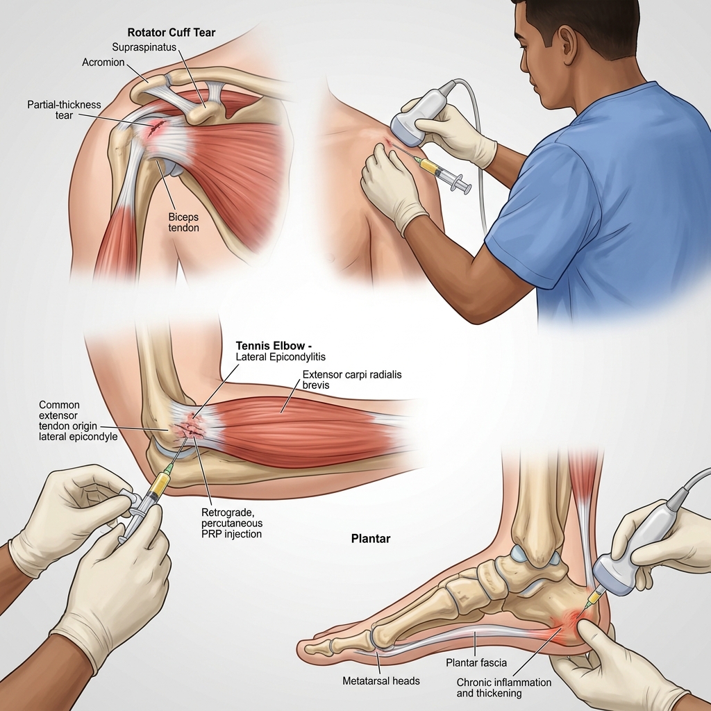
In cases of symptomatic partial-thickness rotator cuff tears, a subacromial or intratendinous injection of PRP forces targeted collagen synthesis to stabilize the frayed tendon fibers before they progress to a full-thickness retraction. I evaluate the exact location of the tear, usually involving the supraspinatus, to determine if the remaining intact fibers can withstand eccentric loading after the biological scaffolding is introduced.
Many patients in Vigan refer to chronic shoulder pain as lamig or ngawit, often seeking traditional hilot massage for relief. While massage may temporarily relieve superficial muscle spasm, applying deep friction to a partially torn rotator cuff tendon can shear the remaining fragile collagen fibers and widen the tear. A clinical assessment is required to differentiate a true rotator cuff tear from cervical radiculopathy (a pinched nerve in the neck), which can mimic shoulder lamig but requires an entirely different treatment pathway. As detailed in our guide on shoulder ultrasound vs MRI for rotator cuff tears, we rely on dynamic ultrasound paired with orthopedic tests to confirm the exact tissue state.
A common assumption is that an MRI showing a rotator cuff tear means surgery is the only option. This is a critical misunderstanding of shoulder biomechanics. Structural imaging only shows tissue anatomy; it does not measure tissue irritability or functional capacity. Many adults have asymptomatic partial tears visible on MRI. If the tear is partial, the tendon fibers are not massively retracted, and the patient maintains basic active range of motion, conservative or regenerative interventions like PRP are highly viable. The clinical decision hinges on the functional deficit, not just the imaging report.
Tennis Elbow (Lateral Epicondylitis): Restoring the ECRB Tendon
Tennis elbow is a degenerative tendinopathy of the extensor carpi radialis brevis (ECRB) tendon at the lateral epicondyle, requiring structural cellular repair rather than simple anti-inflammatory suppression. I target the exact site of ECRB origin with PRP to reverse the degenerative mucinoid changes and restore the tendon's ability to tolerate wrist extension forces during daily activities.
Patients frequently describe this elbow pain as a deep, burning rayuma (rheumatism) that sharply limits their ability to work, grip objects, or play racket sports. True rheumatoid arthritis affects the radiocapitellar joint line with systemic swelling. Lateral epicondylitis is highly localized to the tendon insertion point. When I evaluate this in clinic, I use Cozen's test and resisted wrist extension to isolate the ECRB mechanism and rule out underlying joint pathology.
Internet consensus often suggests that tennis elbow is an inflammatory condition ("itis") that should be aggressively treated with ice and NSAIDs. The physiological reality is that chronic tennis elbow is a failed healing response called angiofibroblastic tendinosis. There are no acute inflammatory cells present after the first few weeks. Aggressively icing the elbow delays the essential infiltration of macrophages needed to clear necrotic tissue.
In my clinic, I look for specific functional deficits before recommending an injection. Furthermore, clinical trials indicate that while steroid injections offer short-term relief, PRP yields statistically significant improvements. At the 6-month mark, PRP patients report a 70% decrease in VAS pain scores, whereas steroid groups often see their pain return to baseline or worse.
Treatment Comparison: PRP vs Corticosteroid
Understanding the long-term impact on your tendons.
PRP treats the structural degradation rather than suppressing a non-existent inflammation (2020; PMID: 26362783).
Plantar Fasciitis: Reversing Plantar Fascia Degeneration
Plantar fasciitis is an overuse degeneration of the plantar fascia insertion at the medial calcaneal tubercle, and injecting PRP directly into this hypovascular insertion site delivers the necessary growth factors to initiate cellular healing. As explained in our clinical guide on morning heel pain and diagnosing plantar fasciitis, my clinical focus is determining if the fascia has thickened on ultrasound, which indicates chronic fibrotic changes that will not resolve with basic stretching alone.
The classic patient presentation is a sharp, stabbing tusok-tusok sensation in the heel during the first steps in the morning. This specific pain pattern occurs because the fascia tightens overnight, and the sudden mechanical load of body weight upon waking micro-tears the tissue anew. It is critical to differentiate this from tarsal tunnel syndrome, where the posterior tibial nerve is compressed. I perform a Tinel's sign test at the ankle and a Windlass test of the toes to confirm whether the pain generator is neurological or structural. If the patient has heel pain accompanied by numbness or tingling radiating into the toes, an entirely different diagnostic pathway is triggered. In conjunction with PRP, offloading via custom foot orthotics and arch support is vital during tissue healing.
Evaluate Heel Pain Accuracy
Rule out nerve entrapment with a clinical assessment in Vigan.
Another persistent myth is that heel spurs cause plantar fasciitis pain. Heel spurs are osteophytes that form as a secondary structural adaptation to chronic tension, not the primary cause of the pain. Surgically removing a heel spur does not fix the underlying fascial degeneration. Pain relief correlates with the healing of the fascial tissue, which is why PRP interventions that stimulate tissue repair often resolve the pain even while the spur remains entirely visible on an X-ray (2020; PMID: 32336895).
The Clinical Reality of PRP Treatment
When I read through patient forums like Reddit or watch comments on YouTube, I constantly see patients frustrated that their tendon pain hasn't vanished after a single PRP shot. The internet often sets unrealistic expectations. Translating regenerative medicine research into actual patient care requires balancing optimal protocols with practical realities. Systematic reviews confirm that PRP provides significant long-term pain relief and functional improvement for chronic tendinopathies when compared to alternative conservative treatments (2019; PMID: 29268037).
The ideal evidence-based protocol involves precise, ultrasound-guided intra-tendinous injections followed by a highly regimented, supervised eccentric loading program to ensure the new collagen fibers align correctly under stress. However, in a provincial setting like Ilocos Sur, patients often face constraints regarding the cost of the PRP preparation kits and the logistical difficulty of attending frequent, supervised physical therapy sessions. Therefore, my approach modifies the ideal standard into a feasible clinical pathway. I ensure the injection is meticulously placed using ultrasound, but I transition the patient to a heavily structured, progressive home-exercise program utilizing isometric loading. I monitor their safety and neurologic status through intermittent telemedicine check-ins and specific in-clinic functional milestones. If a patient experiences resting pain greater than 7/10 or exhibits active infection, they are strictly excluded from the PRP protocol until the acute flare is managed.
The PRP Procedure: What to Expect in Clinic
The clinical administration of PRP is a precise, multi-step process designed to maximize biological yield and ensure accurate delivery.
1 Blood Draw & Centrifugation
A standard phlebotomy procedure collects 15 to 30 milliliters of your whole blood. It is placed in a specialized centrifuge and spun at exact speeds to separate the platelet-rich plasma layer from the red blood cells.
2 Ultrasound-Guided Injection
After applying local anesthesia, I use high-resolution diagnostic ultrasound to visualize the exact location of the tendon tear or fascial thickening, guiding the needle in real-time to deliver the PRP directly into the damaged tissue bed.
The Crucial Role of Rehabilitation After PRP
A biological injection provides the cellular building blocks for repair, but mechanical loading dictates how those blocks are assembled. Without targeted rehabilitation, the newly synthesized collagen will form in a disorganized, chaotic pattern (scar tissue), leaving the tendon weak and prone to re-injury. I require patients to adhere to a strict progression criteria before advancing their activity levels.
Following a PRP procedure, the traditional R.I.C.E. method (Rest, Ice, Compression, Elevation) is strongly contraindicated. Icing the area suppresses the exact inflammatory cascade the PRP was just injected to create. Absolute rest prevents the mechanical signaling (mechanotransduction) required to tell the fibroblasts how to align the new collagen fibers.
Instead, I implement the P.E.A.C.E. & L.O.V.E. protocol paired with early isometric loading. For example, a patient with tennis elbow will begin pain-free isometric wrist extension holds (30 seconds, 4 repetitions) within the first week. Isometric contraction triggers cortical inhibition to reduce local pain (an analgesic effect) and provides a safe mechanical stimulus to the ECRB tendon without introducing the shearing forces of full-range motion. The clinical threshold to progress to heavier eccentric loading is the ability to perform these isometrics completely pain-free. If sharp pain occurs during the isometric phase, the load is immediately reduced to protect the maturing tissue scaffold (Lavallee et al., 2021; PMID: 33727517).
Many patients expect PRP to work like a steroid injection, providing total pain relief within 48 hours. This expectation leads to premature return to sports and rapid re-injury. PRP fundamentally alters tissue architecture, a biological process that takes a minimum of six to twelve weeks to mature. The initial two weeks often involve an increase in localized pain as the inflammatory cascade peaks. True functional recovery is measured in months, marked by a gradual increase in load tolerance and a decrease in morning stiffness, not sudden overnight relief.
Safety, Contraindications, and Next Steps
While PRP utilizes your body's own biological material, minimizing the risk of allergic reaction or systemic side effects, it is not appropriate for every patient.
Critical contraindications
Uncontrolled conditions: Strict exclusion for patients with uncontrolled diabetes mellitus, active systemic infections, or underlying bleeding disorders.
Massive Structural Failure: If ultrasound reveals a massive, full-thickness tendon tear with severe fiber retraction (gap greater than 1.5 cm), PRP will not physically bridge the gap. Surgical referral is required.
If your joint pain is accompanied by systemic fevers (temperature over 38.5°C), sudden profound muscle weakness, or a complete inability to bear weight, do not wait for a regenerative consultation. These are red flags that necessitate immediate evaluation at an emergency department or urgent care facility to rule out septic arthritis or acute total ruptures. If you are struggling with chronic tendon pain that has failed standard rest and physical therapy, a proper diagnostic workup is required to determine if the tissue is a candidate for regenerative repair.
Explore More Regenerative Treatments
This procedure is part of our comprehensive PRP therapy Vigan protocol. Discover how we utilize orthobiologics for natural joint preservation and tissue repair.
Return to Rehabilitation Treatments ViganPlatelet-Rich Plasma (PRP) Realities
Understanding the biological healing timeline of orthobiologics.
Delayed Structural Healing
Chronic tendon injuries rarely heal with complete rest; rest accelerates collagen disorganization.
Collagen Repair
Cortisone degrades tendon fibers; PRP stimulates collagen repair.
Structural Degeneration
Plantar fasciitis is not inflammation; it is structural fascial degeneration.
References & Clinical Evidence
- [1] Arirachakaran A, Sukthuayat A, Sisayanarane T, Laoratanavoraphong S, Kanchanatawan W, Kongtharvonskul J. Platelet-rich plasma versus autologous blood versus steroid injection in lateral epicondylitis: systematic review and network meta-analysis. J Orthop Traumatol. 2016 Jun;17(2):101-12. doi: 10.1007/s10195-015-0376-5. PMID: 26362783.
- [2] Chen X, Jones IA, Park C, Vangsness CT Jr. The Efficacy of Platelet-Rich Plasma on Tendon and Ligament Healing: A Systematic Review and Meta-analysis With Bias Assessment. Am J Sports Med. 2018 Jul;46(8):2020-2032. doi: 10.1177/0363546517743746. PMID: 29268037.
- [3] Mohammed W, Farah S, Nassiri M, McKenna J. Therapeutic efficacy of platelet-rich plasma injection compared to corticosteroid injection in plantar fasciitis: A systematic review and meta-analysis. J Orthop. 2020 Nov-Dec;22:124-134. doi: 10.1016/j.jor.2020.03.053. PMID: 32336895.
- [4] Lavallee M, Bush C. Ultrasound-Guided Percutaneous Tenotomy and Its Associated Pain Reduction and Functionality Outcomes in Nonelite Active Adults. Am J Phys Med Rehabil. 2021 Apr 1;100(4):349-353. doi: 10.1097/PHM.0000000000001570. PMID: 33727517.
* Clinical references are provided to support the medical claims made in this article. TeraCare adheres to evidence-based practices in physical medicine and rehabilitation.

Dr. Ben Rabara
Dr. Ben Rabara is a Board-Certified Physiatrist specializing in Physical Medicine and Rehabilitation. He focuses on non-surgical, precision treatments for musculoskeletal conditions, utilizing advanced diagnostics like MSK Ultrasound.
Medical Disclaimer: The information provided in this article is for educational purposes only and does not substitute for professional medical advice, diagnosis, or treatment. Always consult a qualified physician for your specific health conditions.