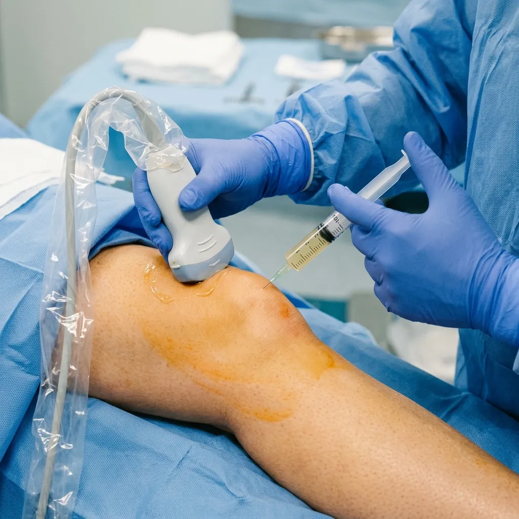
- — Platelet-Rich Plasma (PRP) is an autologous orthobiologic treatment that utilizes your own blood platelets to deliver concentrated growth factors directly to damaged joint tissues.
- — The biological healing process occurs in three distinct phases: the inflammatory phase (1-7 days), the proliferative phase (1-4 weeks), and the remodeling phase (4-12+ weeks).
- — Real, evidence-based PRP focuses on using your own body's active proteins to regulate inflammation and stimulate repair, whereas unproven commercial 'stem cell' tourism carries significant legal, financial, and infection risks.
- — Cartilage has no direct blood supply (avascular), making natural self-repair extremely limited; PRP bypasses this barrier by supplying growth factors directly to the cartilage matrix under ultrasound guidance.
What is Platelet-Rich Plasma? The Autologous Science
Platelet-Rich Plasma (PRP) is an autologous orthobiologic treatment that utilizes a concentrated solution of a patient's own blood platelets suspended in plasma to accelerate tissue healing and modulate chronic inflammation. The term autologous is a medical definition referring to cells, tissues, or proteins obtained from the same individual who receives the treatment. In my clinic, I often explain that PRP is not a drug or a synthetic chemical; rather, it is a highly concentrated dose of your own body's natural healing elements. In the spectrum of physical medicine, we classify PRP as an orthobiologic, which is a substance naturally found in the body that orthopedic specialists and physiatrists use to help bones, joints, muscles, and ligaments heal faster.
To understand the clinical utility of PRP, we must first look at the basic physiology of human blood. Your blood is primarily composed of four main components:
- Red Blood Cells (Erythrocytes): Cells responsible for carrying oxygen throughout the body.
- White Blood Cells (Leukocytes): Immune cells that fight infections and coordinate inflammatory responses.
- Plasma: The liquid base of the blood that carries nutrients, hormones, and proteins.
- Platelets (Thrombocytes): Cell fragments that are best known for clotting blood, but also contain a dense concentration of growth factors.
When you suffer an injury, your body's immediate physiological response is to send platelets to the site of damage. Platelets contain tiny storage sacs called alpha-granules, which house biological proteins known as growth factors. When these platelets arrive at the injured joint or ligament, they release these growth factors to initiate the healing cascade. However, in areas like the joints, tendons, and cartilage, the blood supply is naturally poor, which frequently causes the healing process to stall. PRP resolves this limitation by delivering a concentrated dose of platelets—often 5 to 7 times the baseline concentration found in whole blood—directly into the target area (Cole et al., 2022; PMID: 36410877).
When a patient walks into my clinic, they often present with a complex array of symptoms. They describe a sharp catch when climbing stairs, a constant dull ache at rest, and sometimes a radiating discomfort that travels down their leg. Their joint feels stiff and heavy in the mornings, accompanied by a weak sensation that makes them feel unstable. Some report a warm, burning pain or a tingling numbness around the knee, while others describe a mechanical catching or a loud grinding sound (crepitus) during movement. In my assessment, these symptoms are often the result of progressive cartilage wear or micro-tears in the ligaments. By extracting the patient's own platelets and concentrating them, we can deliver a precise therapeutic payload to kickstart the healing of these damaged structures.
The autologous preparation process is performed directly in our clinic and takes approximately 30 to 45 minutes from start to finish:
- Venipuncture: We draw a small volume of blood (typically 15 to 30 mL) from a vein in your arm, similar to a standard laboratory blood test.
- Centrifugation (The Spinning Process): The blood is placed in a specialized sterile vial and spun in a high-speed centrifuge. This process separates the blood components based on their molecular weight. A first spin separates the red blood cells from the plasma and platelets. A second spin (the double-spin technique) further concentrates the platelets into a dense pellet.
- Extraction and Activation: We extract the platelet-rich fraction, separating it from the platelet-poor plasma and red blood cell layers. In my practice, depending on the tissue state and specific pathology, we may activate the platelets using calcium chloride to trigger the immediate release of growth factors prior to injection.
How PRP Stimulates Cartilage Repair at the Cellular Level
Articular cartilage is the smooth, slippery tissue covering the ends of bones inside a joint, allowing them to glide over one another without friction. However, cartilage is avascular, meaning it has no direct blood supply, and it is aneural, meaning it contains no nerves. Because it lacks a blood supply, cartilage has an extremely limited capacity for self-repair. When cartilage is damaged due to wear-and-tear or injury, it cannot pull healing cells from the bloodstream. This is where Platelet-Rich Plasma provides a critical biological intervention (Li M et al., 2022; PMID: 36086807). Often, cartilage wear leads to chronic fluid buildup; this joint effusion is a medical term for the excess fluid accumulation that causes grinding and stiffness in the knee joint.
When we inject concentrated platelets directly into a damaged joint, the platelets undergo a process called degranulation, releasing a massive wave of growth factors and cytokines from their alpha-granules. These proteins do not act as structural replacements for cartilage. Instead, they act as cellular messengers, binding to specific receptors on the surface of local cells to initiate a cascade of tissue regeneration. In my clinical reasoning, understanding which growth factors are interacting is vital for predicting how a joint will respond to therapy (Andia & Maffulli, 2016; PMID: 27109909):
- Platelet-Derived Growth Factor (PDGF): Stimulates cell replication, increases collagen synthesis, and recruits local stem cells to the site of damage.
- Transforming Growth Factor-Beta (TGF-β): Encourages chondrocytes (cartilage-producing cells) to multiply and increases the production of extracellular matrix components, specifically collagen type II and proteoglycans.
- Insulin-like Growth Factor-1 (IGF-1): Protects chondrocytes from programmed cell death (apoptosis) and coordinates the synthesis of the cartilage matrix.
- Vascular Endothelial Growth Factor (VEGF): Stimulates angiogenesis (the formation of new blood vessels) in surrounding tissues, bringing vital oxygen and nutrients to support the remodeling process.
This biological influx accomplishes two primary goals: it shifts the joint chemistry from a catabolic (tissue-destroying) state to an anabolic (tissue-building) state, and it significantly downregulates chronic inflammatory cytokines like Interleukin-1 (IL-1) and Tumor Necrosis Factor-alpha (TNF-α). Multiple clinical studies, including systematic reviews and randomized controlled trials, have confirmed that PRP injections reduce joint pain, improve function, and support cartilage health in patients with mild-to-moderate knee osteoarthritis (Tan J et al., 2021; PMID: 33429777; Meng F et al., 2023; PMID: 38037038). The goal of PRP is joint preservation—prolonging the life of your natural joint and delaying or preventing the need for a surgical replacement.
Differentiating Real PRP from Shady 'Stem Cell' Tourism
In my practice, I frequently review discussions on online patient forums such as Reddit, YouTube comments, and health communities like patientslikeme and inspire. A common trend I observe is that patients are often confused by the differences between Platelet-Rich Plasma (PRP) and "stem cell therapy." Because both are marketed under the umbrella of "regenerative medicine," patients frequently assume they are interchangeable. This confusion is actively exploited by commercial clinics offering unproven, highly expensive procedures. It is essential to differentiate evidence-based, autologous PRP from commercialized stem cell tourism.
Standard PRP is a heavily researched, highly regulated, and clinically proven autologous treatment. Because it utilizes your own platelets to stimulate your body's existing cells, it carries minimal risk and is prepared transparently in a clinical setting. In contrast, "stem cell tourism" refers to the practice of traveling to offshore or unregulated clinics that advertise miracle cures using imported, commercially prepared stem cell products (such as amniotic, placental, or umbilical cord tissue). Many of these clinics make bold, scientifically unsupported claims that their injections can completely regrow bone-on-bone joints or cure systemic diseases (Naderi N et al., 2020; PMID: 31930387).
In my assessment, patients must be aware of the following medical and scientific realities regarding commercial stem cell claims:
- The "Dead Cell" Reality: Laboratory analyses of commercially available amniotic and umbilical cord products have repeatedly shown that these products contain zero live stem cells after the freezing, sterilization, and distribution processes. Patients are paying thousands of dollars for expensive injections of dead proteins, not live cells.
- Safety and Infection Risks: Because these commercial products are derived from donor tissues rather than the patient's own body, they carry risks of tissue rejection, severe inflammatory reactions, and transmission of infectious diseases. Furthermore, unregulated offshore processing facilities have been linked to severe bacterial joint infections.
- Lack of Clinical Standardization: Unlike PRP, which has standardized preparation protocols and extensive clinical trials backing its use for joint pain, many commercial stem cell injections lack standardized dosing, clinical guidelines, or long-term safety data.
I recommend that patients focus on established, evidence-based orthobiologics. Autologous PRP therapy utilizes your body's own active proteins to modulate joint inflammation and protect cartilage safely, without the extreme financial exploitation and safety risks associated with unregulated stem cell advertising (Meng F et al., 2023; PMID: 38037038).
The Necessity of Dynamic Ultrasound-Guided Injections
The success of Platelet-Rich Plasma therapy depends entirely on diagnostic and placement accuracy. In my clinical experience, performing a joint injection "blindly"—relying only on physical touch and anatomical landmarks on the skin—is an obsolete standard that compromises patient outcomes. Research shows that up to 20% to 30% of blind knee injections fail to deliver the therapeutic payload into the joint capsule, instead placing the platelets into the surrounding fat pad, muscle, or subcutaneous tissues where they cannot interact with the cartilage matrix.
To eliminate this margin of error, I perform every PRP injection under high-resolution, live musculoskeletal ultrasound guidance. An ultrasound is a dynamic diagnostic imaging modality that uses high-frequency sound waves to visualize tissues in active motion. In contrast, an MRI is a static imaging scan that uses magnetic fields to capture snapshots of the joint space. This approach provides several key clinical benefits:
- Real-Time Visualization: I scan the joint in motion to identify the exact pocket of cartilage wear, joint effusion, or ligament fraying. We see the needle tip enter the target structure in real-time, ensuring 100% placement accuracy.
- Avoiding Critical Structures: Ultrasound guidance allows us to avoid blood vessels, nerves, and healthy tendons, reducing post-injection pain and avoiding structural damage.
- Dynamic Assessment: Unlike static MRI scans, ultrasound allows us to move the joint during the scan. This dynamic assessment helps identify impingements or instability that only appear when the joint is in motion.
By combining high-concentration PRP with dynamic ultrasound guidance, we ensure that the healing factors are deposited precisely where they are needed, maximizing the biological stimulus for cartilage repair and joint stabilization.
Rehabilitation Pathway and Local Realities in the Philippines
A common medical myth is that a PRP injection is a passive cure—that you can receive the shot, rest on the couch, and expect your joint to heal completely. In my practice, I emphasize that biological injections are only the first half of joint preservation. The second half is progressive mechanical loading. The concentrated platelets deliver the growth factors to initiate repair, but these new, delicate tissue fibers require controlled movement and mechanical stress to align properly and mature into strong, functional collagen scaffolds. Physical rehabilitation is the mandatory partner to PRP.
To help patients visualize their options, the table below compares evidence-based PRP, standard cortisone steroids, and commercial stem cell tourism across critical clinical and financial parameters:
| Parameters | Cortisone Steroids | Evidence-Based PRP | Stem Cell Tourism |
|---|---|---|---|
| Primary Mechanism | Chemical suppression of inflammation (temporary pain mask) | Biological modulation and cartilage matrix stimulation (repair) | Unproven claims of complete tissue regeneration using donor tissues |
| Cartilage Impact | Chondrotoxic (accelerates cartilage wear if repeated) | Chondroprotective (protects cartilage and delays degeneration) | Unverified (often injects dead proteins with no cell viability) |
| Preparation Type | Synthetic pharmaceutical | Autologous (100% your own blood) | Allo-transplant (donor tissues with rejection/infection risks) |
| Efficacy Duration | Short-term (4 to 12 weeks of temporary relief) | Long-term (12+ months of functional improvement) | Unproven (lacks consistent clinical trials or guidelines) |
| Relative Cost | Low initial cost | Moderate (highly cost-efficient compared to surgery) | Extremely High (often thousands of dollars out-of-pocket) |
A typical cartilage and joint rehabilitation plan at our Vigan City clinic follows a structured, three-phase care pathway adapted to the physiological stages of tissue repair:
Phase 1: The Inflammatory Phase (Days 1 to 7)
The goal is to protect the joint and allow the concentrated platelets to trigger the initial repair cascade.
- Biological Activity: Platelets degranulate, releasing growth factors and initiating a controlled inflammatory response to recruit healing cells.
- Timelines & Rest: Restrict high-impact activities, heavy lifting, or prolonged standing for the first 48 to 72 hours. Gentle walking around the home is encouraged, but strenuous exercise must be avoided.
- Friction Alert (No NSAIDs): Because PRP relies on a localized inflammatory response to jumpstart healing, patients must avoid taking anti-inflammatory pain relievers (NSAIDs like Ibuprofen, Naproxen, or Mefenamic Acid) for at least 2 weeks after PRP. These drugs block the inflammatory cascade, neutralizing the injection's benefits. We manage pain using ice and acetaminophen.
- Warning Signs (Red Flags): If you experience a high fever, chills, rapidly spreading redness around the joint, or severe throbbing pain unresponsive to acetaminophen, contact the clinic immediately to rule out infection.
Phase 2: The Proliferation Phase (Weeks 2 to 6)
The goal is to introduce controlled mechanical loading to guide the alignment of new collagen fibers.
- Biological Activity: Fibroblasts lay down new collagen type I and III fibers, creating new tissue scaffolding.
- Clinical Rehabilitation: Begin structured physical therapy. We focus on low-load, isometric exercises (isometric quadriceps sets, straight leg raises) and range-of-motion mobilizations. This strengthens the surrounding muscles—the primary shock absorbers of the joint—without putting shearing stress on the healing cartilage. Doing 3 sets of 10 to 12 repetitions is standard practice.
- Timelines: Exercise frequency is set to 3 to 4 sessions per week, keeping pain below a 3 out of 10 threshold.
Phase 3: The Remodeling Phase (Weeks 7 to 12+)
The goal is to build dynamic stability, restore full range of motion, and transition to independent function.
- Biological Activity: Collagen fibers mature, cross-link, and align along the lines of mechanical stress, significantly increasing tissue tensile strength.
- Clinical Rehabilitation: Transition to progressive loading exercises. This includes closed-kinetic chain exercises (wall sits, leg presses, step-downs) and balance/proprioception training. We teach the joint to adapt to dynamic loads in motion.
- Home Program Stop Rules: If any exercise causes swelling or pain that persists for more than 24 hours, the patient must stop that exercise, return to the previous loading level, and consult our rehab team.
In Northern Luzon, we adapt this pathway to realistic local conditions. Many of our patients in Ilocos Sur and nearby provinces travel from farming municipalities or outlying towns (such as Bantay, San Ildefonso, Santo Domingo, or Santa Maria). Requiring them to travel to Vigan City daily for supervised physical therapy is practically unfeasible and financially draining. To bridge this gap, we perform the precise biological injections in our clinic, guide the patient through their initial exercises, and construct a highly detailed, home-based progressive loading guide. We follow up during their scheduled injection visits (typically a series of 2 to 3 sessions spaced 3 to 4 weeks apart) or via telehealth to monitor their progression and adjust loading parameters safely.
Furthermore, we strongly advise patients against using traditional hilot (aggressive massage) on a joint that has recently received PRP. While gentle topical application of oil is harmless, aggressive rubbing or pressing on the joint capsule can displace the injected platelets, disrupt the localized inflammatory cascade required for healing, and damage the newly forming collagen scaffolds, neutralizing the benefits of the procedure.
References & Clinical Evidence
- [1] Li M, et al. Platelet Rich Plasma in the Repair of Articular Cartilage Injury: A Narrative Review. Front Bioeng Biotechnol. 2022 Sep;10:9465610. doi: 10.3389/fbioe.2022.9465610. PMID: 36086807.
- [2] Chen X, et al. Platelet-rich plasma treatment for talar cartilage repair: a systematic review and meta-analysis. J Orthop Surg Res. 2023 May 13;18(1):354. doi: 10.1186/s13018-023-03822-9. PMID: 37161527.
- [3] Tan J, et al. Effects of platelet-rich plasma injection for pain control and cartilage clinical outcomes in knee osteoarthritis: a systematic review and meta-analysis. J Orthop Surg Res. 2021 Jan;16(1):29. doi: 10.1186/s13018-020-02187-5. PMID: 33429777.
- [4] El-Kadiry AE, et al. Double-Blind Randomized Controlled Trial Comparing Platelet-Rich Plasma With Intra-Articular Corticosteroid Injections in Patients With Bilateral Knee Osteoarthritis. Cureus. 2022 Oct;14(10):e30362. doi: 10.7759/cureus.30362. PMID: 36324362.
- [5] Meng F, et al. Efficacy and safety of corticosteroids, hyaluronic acid, and PRP and combination therapy for knee osteoarthritis: a systematic review and network meta-analysis. Medicine (Baltimore). 2023 Dec 1;102(48):e38038. doi: 10.1097/MD.0000000000038038. PMID: 38037038.
- [6] Naderi N, et al. Regenerative Medicine, Stem Cell Therapies, and Platelet-Rich Plasma: Where Is the Evidence? Aesthet Surg J. 2020 Mar;40(4):460-465. doi: 10.1093/asj/sjz387. PMID: 31930387.
- [7] HTA Report. Platelet-Rich Plasma (PRP) for Knee Osteoarthritis Technology Overview. CADTH Health Technology Review. 2024 Jan. PMID: 38295392.
* Clinical references are provided to support the medical claims made in this article. TeraCare adheres to evidence-based practices in physical medicine and rehabilitation.

Dr. Ben Rabara
Dr. Ben Rabara is a Board-Certified Physiatrist specializing in Physical Medicine and Rehabilitation. He focuses on non-surgical, precision treatments for musculoskeletal conditions, utilizing advanced diagnostics like MSK Ultrasound.
Medical Disclaimer: The information provided in this article is for educational purposes only and does not substitute for professional medical advice, diagnosis, or treatment. Always consult a qualified physician for your specific health conditions.